Apply the rules of medical language to build, analyze, spell, pronounce, abbreviate, and define terms as they relate to the blood
Identify meanings of keyword components of the blood
Use terms related to the blood
Blood Vessels and Blood Word Parts
Click on prefixes, combining forms, and suffixes to reveal a list of word parts to memorize for the Cardiovascular System – Blood.
Introduction to the Blood Vessels and Blood
Our large, complex bodies need blood to deliver nutrients to and remove wastes from our trillions of cells. The heart, as discussed in the previous chapter, pumps blood throughout the body in a network of blood vessels. Together, these three components—blood, heart, and vessels—makes up the cardiovascular system.
Virtually every cell, tissue, organ, and system in the body is impacted by the circulatory system. This includes the generalized and more specialized functions of transport of materials, capillary exchange, maintaining health by transporting white blood cells and various immunoglobulins (antibodies), hemostasis, regulation of body temperature, and helping to maintain acid-base balance. Table 7.1 summarizes the important relationships between the circulatory system and the other body systems.
Table 7.1 Interaction of the Circulatory System with Other Body Systems. A table depicting the various body systems and the role of the circulatory system in each. Adapted from Betts, et al., 2021. Licensed under CC BY 4.0.
SYSTEM
ROLE OF CIRCULATORY SYSTEM
DigestiveDigestive System
Absorbs nutrients and water; delivers nutrients (except most lipids) to liver for processing by hepatic portal vein; provides nutrients essential for hematopoiesis and building hemoglobin.
EndocrineEndocrine System
Delivers hormones: atrial natriuretic hormone (peptide) secreted by the heart atrial cells to help regulate blood volumes and pressures; epinephrine, ANH, angiotensin II, ADH, and thyroxine to help regulate blood pressure; estrogen to promote vascular health in women and men.
IntegumentaryIntegumentary System
Carries clotting factors, platelets, and white blood cells for hemostasis, fighting infection, and repairing damage; regulates temperature by controlling blood flow to the surface, where heat can be dissipated; provides some coloration of integument; acts as a blood reservoir.
LymphaticLymphatic System
Transports various white blood cells, including those produced by lymphatic tissue, and immunoglobulins (antibodies) throughout the body to maintain health; carries excess tissue fluid not able to be reabsorbed by the vascular capillaries back to the lymphatic system for processing.
MuscularMuscular System
Provides nutrients and oxygen for contraction; removes lactic acid and distributes heat generated by contraction; muscular pumps aid in venous return; exercise contributes to cardiovascular health and helps to prevent atherosclerosis.
NervousNervous System
Produces cerebrospinal fluid (CSF) within choroid plexuses; contributes to blood-brain barrier; cardiac and vasomotor centers regulate cardiac output and blood flow through vessels via the autonomic system.
ReproductiveReproductive System
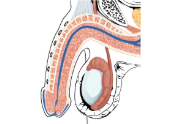
Aids in erection of genitalia in both sexes during sexual arousal; transports gonadotropic hormones that regulate reproductive functions.
RespiratoryRespiratory System
Provides blood for critical exchange of gases to carry oxygen needed for metabolic reactions and carbon dioxide generated as byproducts of these processes.
SkeletalSkeletal System
Provides calcium, phosphate, and other minerals critical for bone matrix; transports hormones regulating buildup and absorption of matrix including growth hormone (somatotropin), thyroid hormone, calcitronins, and parathyroid hormones; erythropoietin stimulates myeloid cell hematopoiesis; some level of protection for select vessels by bony structures.
UrinaryUrinary System
Delivers 20% of resting circulation to kidneys for filtering, reabsorption of useful products, and secretion of excesses; regulates blood volume and pressure by regulating fluid loss in the form of urine and by releasing the enzyme renin that is essential in the renin-angiotensin-aldosterone mechanism.
Cardiovascular System – Blood Vessels and Blood Medical Terms
Anatomy of the Blood Vessels
Blood pumped by the heart flows through a series of vessels known as arteries, arterioles, capillaries, venules, and veins before returning to the heart.
Arteries transport blood away from the heart and branch into smaller vessels, forming arterioles.
Arterioles distribute blood to capillary beds, the sites of exchange with the body tissues.
A capillary is a microscopic channel that supplies blood to the tissues themselves, a process called perfusion.
Exchange of gases and other substances occurs in the capillaries between the blood and the surrounding cells and their tissue fluid (interstitial fluid).
For capillaries to function, their walls must be leaky, allowing substances to pass through.
Capillaries lead back to small vessels known as venules.
Venules are small veins that converge into larger veins.
A vein is a blood vessel that conducts blood toward the heart
Compared to arteries, veins are thin-walled vessels with large and irregular lumens
Larger veins are commonly equipped with valves that promote the unidirectional flow of blood toward the heart and prevent backflow toward the capillaries caused by the inherent low blood pressure in veins as well as the pull of gravity
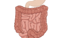
Other ways in which the body assists the transport of venous blood back to the heart involve contractions of skeletal muscles in the extremities (see figure below), as well as pressure variations caused by breathing motion in the chest.
Concept Check
Both arteries and veins have the same three distinct tissue layers, called tunics, for the garments first worn by ancient Romans. From the most interior layer to the outer, these tunics are the tunica intima, the tunica media, and the tunica externa. The smooth muscle in the middle layer, the tunica media, provides the vessel with the ability to vasoconstrict and vasodilate as needed to ensure sufficient blood flow. The table below compares the features of arteries and veins.
Table 7.2. Comparison of Arteries and Veins. From Betts, et al., 2021. Licensed under CC BY 4.0.
CHARACTERISTIC
ARTERIES
VEINS
Direction of blood flow
Conducts blood away from the heart
Conducts blood toward the heart
General appearance
Rounded
Irregular, often collapsed
Pressure
High
Low
Wall thickness
Thick
Thin
Relative oxygen concentration
Higher in systemic arteries
Lower in pulmonary arteries
Lower in systemic veins
Higher in pulmonary veins
Valves
Not present
Present most commonly in limbs and in veins inferior to the heart
The Major Arteries and Veins in the Human Body
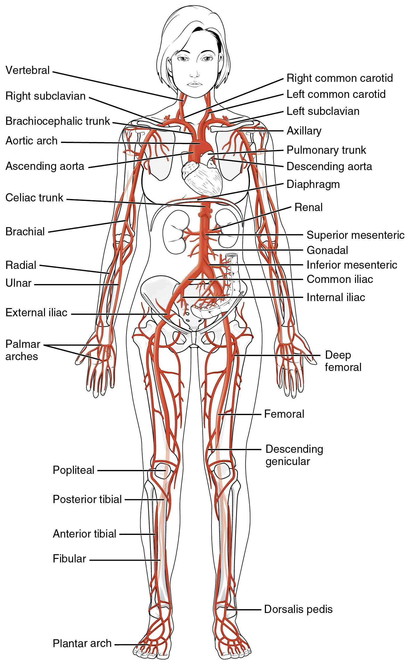
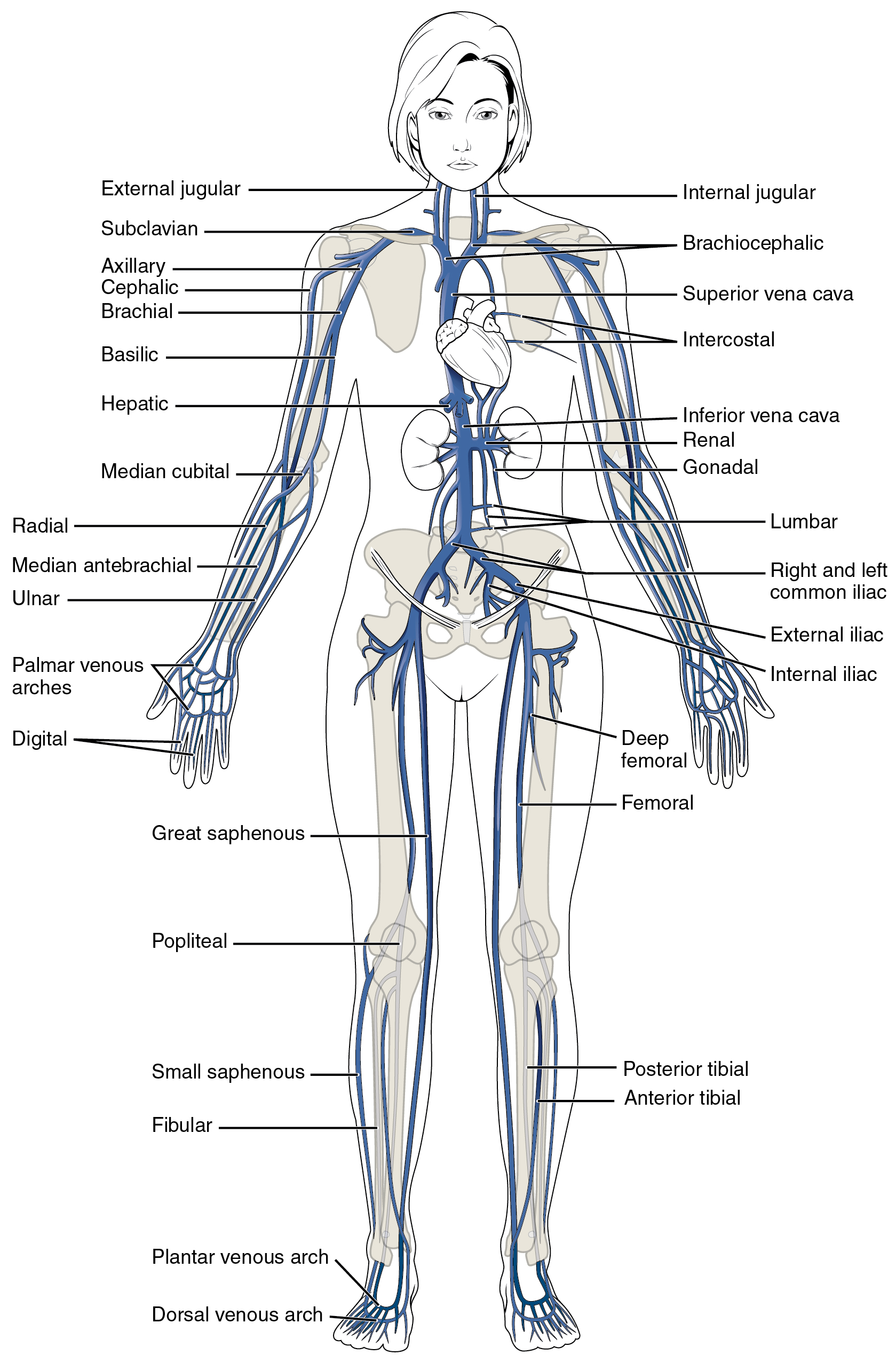
Many arteries and veins share the same names, parallel one another throughout the body, and are very similar on the right and left sides of the body. For example, you will find a pair of femoral arteries and a pair of femoral veins, with one vessel on each side of the body. In contrast, some vessels closer to the midline of the body, such as the aorta, are unique and not paired. Names of vessels may change with location. Like a street that changes name as it passes through an intersection, an artery or vein can change names as it passes an anatomical landmark. For example, the left subclavian artery becomes the axillary artery as it passes into the axillary region, and then becomes the brachial artery as it enters the upper arm. The next two diagrams illustrate the major arteries and veins in the human body.
Figure 7.1 Systemic Arteries. The major systemic arteries shown here deliver oxygenated blood throughout the body. From Betts, et al., 2021. Licensed under CC BY 4.0.Figure 7.2 Major Systemic Veins of the Body. The major systemic veins of the body are shown here in an anterior view. From Betts, et al., 2021. Licensed under CC BY 4.0.
Concept Check
Without looking back at the images of the main arteries and veins of the body, can you name and locate 3 arteries and 3 veins in your body?
Physiology of the Blood Vessels
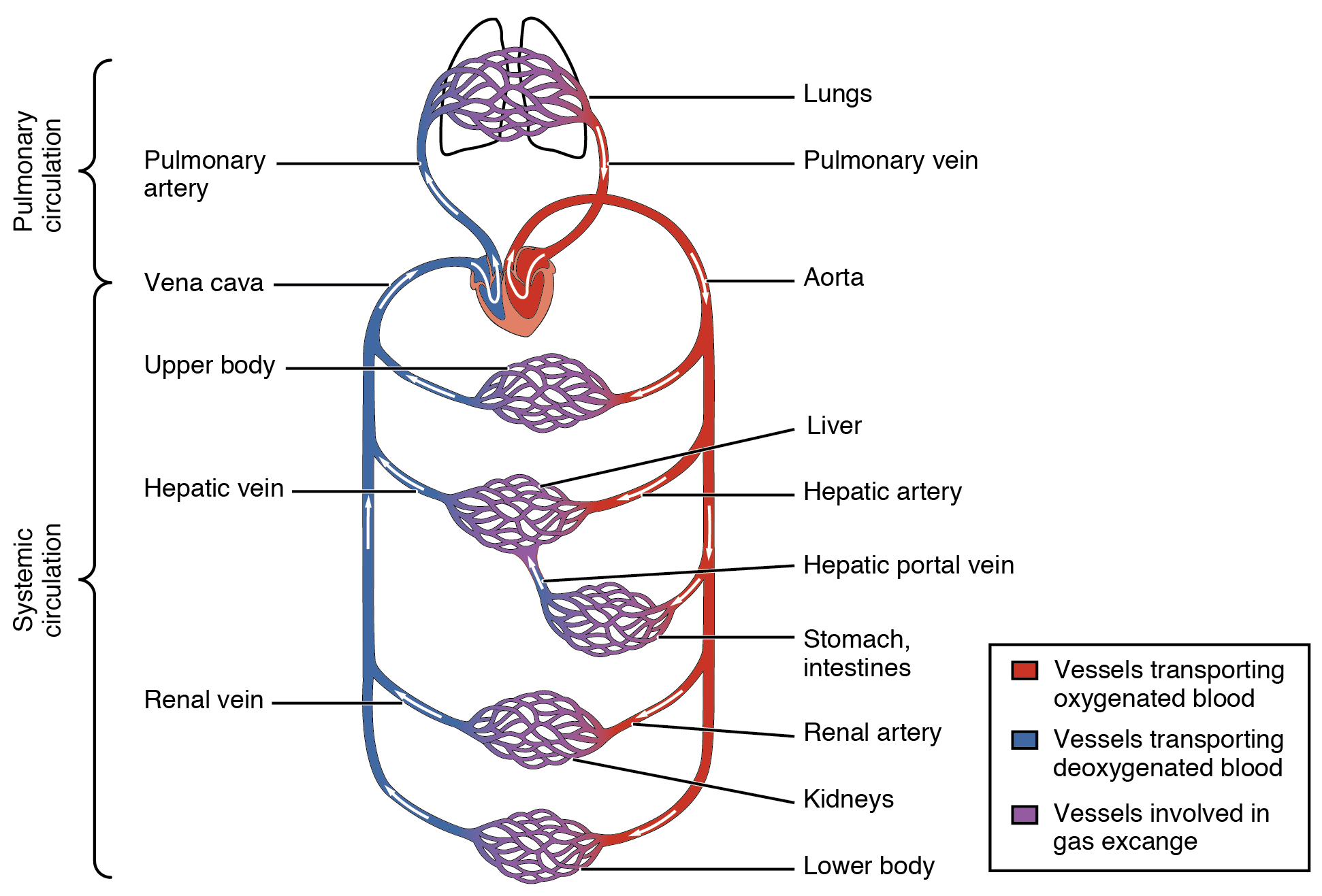
Arteries and veins transport blood in two distinct circuits: the systemic circuit and the pulmonary circuit. Systemic arteries provide blood rich in oxygen to the body’s tissues. The blood returned to the heart through systemic veins has less oxygen, since much of the oxygen carried by the arteries has been delivered to the cells. In contrast, in the pulmonary circuit, arteries carry blood low in oxygen exclusively to the lungs for gas exchange. Pulmonary veins then return freshly oxygenated blood from the lungs to the heart to be pumped back out into systemic circulation.
Figure 7.3 Cardiovascular Circulation. The pulmonary circuit moves blood from the right side of the heart to the lungs and back to the heart. The systemic circuit moves blood from the left side of the heart to the head and body and returns it to the right side of the heart to repeat the cycle. The arrows indicate the direction of blood flow, and the colors show the relative levels of oxygen concentration. From Betts, et al., 2021. Licensed under CC BY 4.0.
Blood Pressure
Blood pressure is the force exerted by blood upon the walls of the blood vessels or the chambers of the heart. Blood pressure may be measured in capillaries and veins, as well as the vessels of the pulmonary circulation; however, the general term ‘blood pressure’ refers to the pressure of blood flowing in the arteries of the systemic circulation. Blood pressure is one of the critical parameters measured on virtually every patient in every healthcare setting. The technique used today was developed more than 100 years ago by a pioneering Russian physician, Dr. Nikolai Korotkoff. Turbulent blood flow through the vessels can be heard as a soft ticking while measuring blood pressure; these sounds are known as Korotkoff sounds. Blood pressure is measured in mm Hg and is usually obtained from the brachial artery using a sphygmomanometer and a stethoscope. Blood pressure is recorded as systolic pressure over diastolic pressure.
Did You Know?
120/80 mm Hg is a normal, healthy blood pressure. 60-100 beats per minute is a normal, resting, adult pulse.
Five variables influence blood flow and blood pressure:
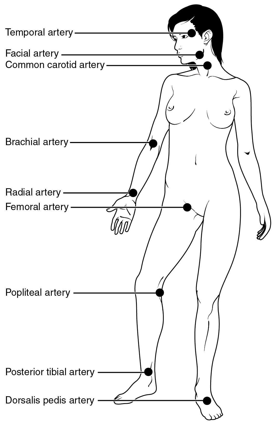
Each time the heart ejects blood forcefully into the circulation, the arteries must expand and then recoil to accommodate the surge of blood moving through them. This expansion and recoiling of the arterial wall is called the pulse and allows us to measure heart rate. Pulse can be palpated manually by placing the tips of the fingers across an artery that runs close to the body surface, such as the radial artery or the common carotid artery. These sites and other pulse sites are shown in the figure below.
Both the rate and the strength of the pulse are important clinically. A high or irregular pulse rate can be caused by physical activity or other temporary factors, but it may also indicate a heart condition. The pulse strength indicates the strength of ventricular contraction and cardiac output. If the pulse is strong, then systolic pressure is high. If it is weak, systolic pressure has fallen, and medical intervention may be warranted.
Figure 7.4 Pulse Sites. The pulse is most readily measured at the radial artery, but can be measured at any of the pulse points shown. From Betts, et al., 2021. Licensed under CC BY 4.0.
The Composition (Anatomy) of Blood and the Functions of the Components
Blood is a connective tissue made up of cellular elements and an extracellular matrix. The cellular elements are referred to as the formed elements and include red blood cells (RBCs), white blood cells (WBCs), and platelets. The extracellular matrix, called plasma, makes blood unique among connective tissues because it is fluid. This fluid, which is mostly water, perpetually suspends the formed elements and enables them to circulate throughout the body within the cardiovascular system.
Did You Know?
Blood constitutes approximately 8% of adult body weight.
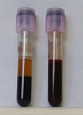
In the laboratory, blood samples are often centrifuged in order to separate the components of blood from one another (see the figure below). Erythrocytes are the heaviest elements in blood and settle at the very bottom of the tube. Above the erythrocyte layer we see the buffy coat, a pale, thin layer of leukocytes and thrombocytes, which together make up less than 1% of the sample of whole blood. Above the buffy coat is the blood plasma, normally a pale, straw-colored fluid, which constitutes the remainder of the sample.
In normal blood, about 45 percent of a sample is erythrocytes, which is referred to as the hematocrit. The hematocrit of any one sample can vary significantly, however, about 36–50 percent, according to gender and other factors. Not counting the buffy coat, which makes up less than 1% of the blood, we can estimate the mean plasma percentage to be the percent of blood that is not erythrocytes: approximately 55%.
Figure 7.5 Composition of Blood: Two tubes of EDTA-anticoagulated blood. Left tube: after standing, the RBCs have settled at the bottom of the tube. Reused from Libretext Anatomy & Physiology
Blood Plasma
Like other fluids in the body, plasma is composed primarily of water. In fact, it is about 92% water. Dissolved or suspended within this water is a mixture of substances, most of which are proteins. The major components of plasma and their functions are summarized in the table below.
Formed Elements (Erythrocytes, Leukocytes, Thrombocytes)
The table below summarizes the main facts about the formed elements in blood.
Table 7.3 Summary of Formed Elements in Blood. Adapted from Betts, et al., 2021.
Licensed under CC BY 4.0.
FORMED ELEMENT
MAJOR SUBTYPES
NUMBER PRESENT PER MICROLITER (µL)AND MEAN (RANGE)
APPEARANCE IN A STANDARD BLOOD SMEAR
SUMMARY OF FUNCTIONS
COMMENTS
Erythrocytes (red blood cells)
Red Blood Cell
n/a
5.2 million ( 4.4-5.0 million)
Flattened biconcave disk; no nucleus; pale red colour
Transport oxygen and some carbon dioxide between tissues and lungs
Lifespan of approximately 120 days
Leukocytes (white blood cells)
n/a
7000 (5000 – 10,000)
Obvious dark-staining nucleus
All function in body defenses
Exit capillaries and move into tissues; lifespan of usually a few hours or days
Leukocytes (white blood cells) Types
Granulocytes including neutrophils, eosinophils, and basophils
4360 (1800-9950)
Abundant granules in cytoplasm; nucleus normal lobed
Nonspecific (innate) resistance to disease
Classified according to membrane-bound granules in cytoplasm
Neutrophils
Neutrophil Cell
4150 (1800-7300)
Nuclear lobes increase with age; pale lilac granules
Phagocytic; particularly effective against bacteria. Release cytotoxic chemicals from granules
Most common leukocyte; lifespan of minutes to days
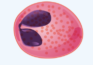
Eosinophils
Eosinophils Cell
165 (0-700)
Nucleus generally two-lobed; bright red-orange granules
Phagocytic cells; particularly effective with antigen-antibody complexes. Release antihistamines. Increase in allergies and parasitic infections
Lifespan of minutes to days
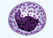
Basophils
Basophil Cell
44 (0-150)
Nucleus generally two-lobed but difficult to see due to presence of heavy, dense, dark purple granules
Promotes inflammation
Least common leukocyte; lifespan unknown
Agranulocytes including lymphocytes and monocytes
2640 (1700-4950)
Lack abundant granules in cytoplasm; have a simple-shaped nucleus that may be indented
Body defenses
Group consists of two major cell types from different lineages
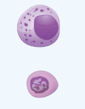
Lymphocytes
Lymphocytes Cell
2185 (1500-4000)
Spherical cells with a single often large nucleus occupying much of the cell’s volume; stains purple; see in large (natural killer cells) and small (B and T cells) variants
Primarily specific (adaptive) immunity; T cells directly attack other cells (cellular immunity). B cells release antibodies (humoral immunity); natural killer cells are similar to T cells but nonspecific
Initial cells originate in bone marrow, but secondary production occurs in lymphatic tissue; several distinct subtypes; memory cells form after exposure to a pathogen and rapidly increase responses to subsequent exposure; lifespan of many years
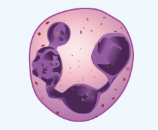
Monocytes
Monocytes Cell
455 (200-950)
Largest leukocyte with an indented or horseshoe-shaped nucleus
Very effective phagocytic cells engulfing pathogens or worn out cells; also serve as antigen-presenting cells (APCs) for other components of the immune system
Produced in red bone marrow; referred to as macrophages after leaving circulation
Platelets
Platelete Cells
n/a
350,000 (150,000 – 500,000)
Cellular fragments surrounded by a plasma membrane and containing granules; purple stain
Hemostasis plus release growth factors for repair and healing of tissue
Formed from megakaryocytes that remain in the red bone marrow and shed platelets into circulation
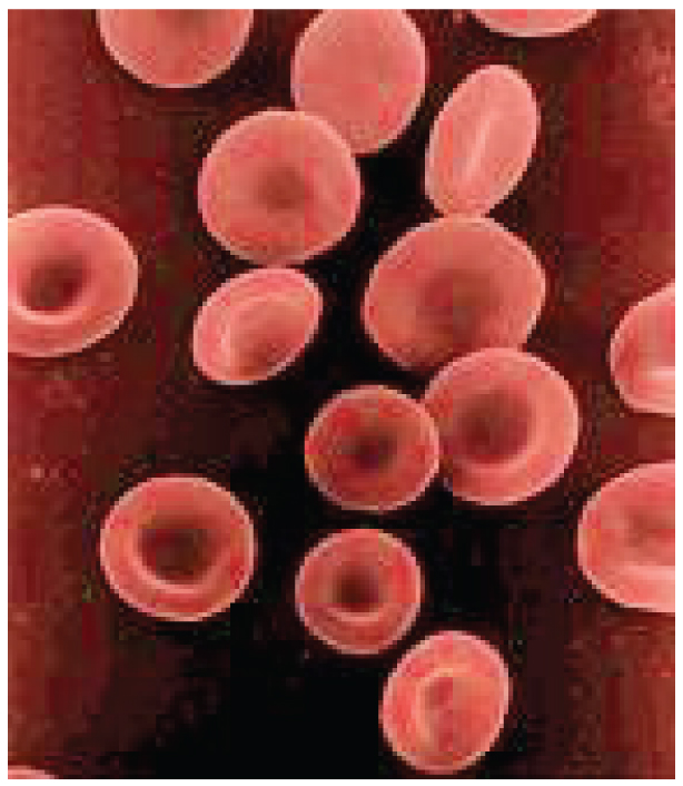
Erythrocytes
The most abundant formed elements in blood, erythrocytes are basically sacs packed with an oxygen-carrying compound called hemoglobin. Production of erythrocytes in the red bone marrow occurs at the staggering rate of more than 2 million cells per second. For this production to occur, raw materials including iron, copper, zinc B-vitamins, glucose, lipids, and amino acids must be present in adequate amounts. Erythrocytes live only 120 days on average, and thus must be continually replaced. Worn-out erythrocytes are phagocytized by macrophages and their hemoglobin is broken down. The breakdown products are recycled or removed as wastes.
Figure 7.6 Shape of Red Blood Cells. Erythrocytes are biconcave discs with very shallow centers. This shape optimizes the ratio of surface area to volume, facilitating gas exchange. It also enables them to fold up as they move through narrow blood vessels. From Betts, et al., 2021. Licensed under CC BY 4.0.
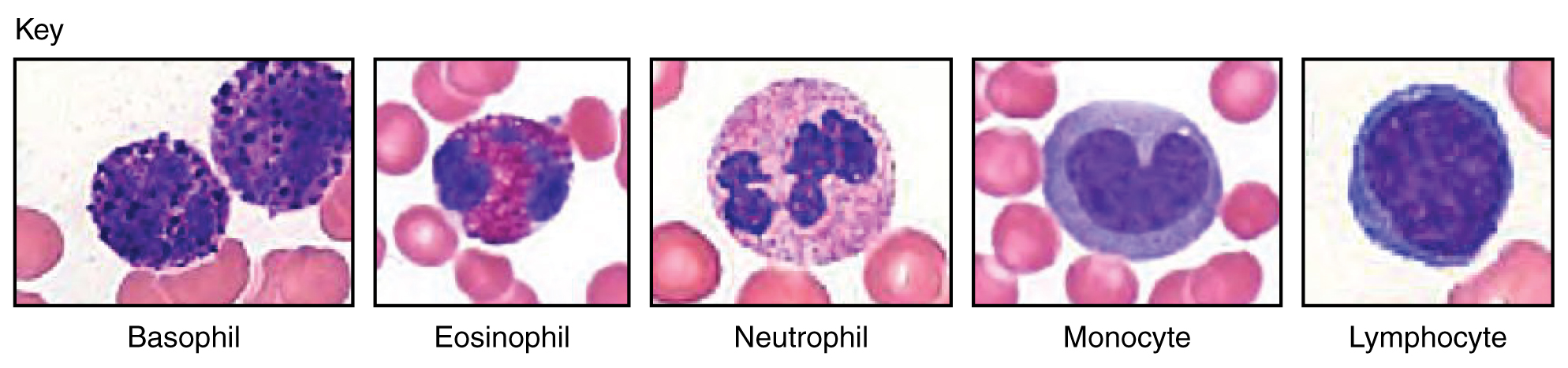
Leukocytes
Leukocytes protect the body against invading microorganisms and body cells with mutated DNA, and they clean up debris, thus they are a major component of the body’s defenses against disease. Figure 7.7 shows the different types of leukocytes. Leukocytes routinely leave the bloodstream to perform their defensive functions in the body’s tissues, where they are often given distinct names, such as macrophage or microglia, depending on their function.
Lymphocytes are one of the types of leukocytes and will be discussed in more detail here, since they tie into the next chapter which discusses the body’s defenses The three major groups of lymphocytes include natural killer cells, B cells, and T cells.
Natural killer (NK) cells are capable of recognizing cells that do not express “self” proteins on their plasma membrane or that contain foreign or abnormal markers. These “nonself” cells include cancer cells, cells infected with a virus, and other cells with atypical surface proteins.
B lymphocytes (B cells) and T lymphocytes (T cells), play prominent roles in defending the body against specific pathogens (disease-causing microorganisms) and are involved in specific immunity. B cells undergo a maturation process in the bone marrow, whereas T cells undergo maturation in the thymus. This site of the maturation process gives rise to the name B and T cells.
Plasma cells, a type of B cell, produce the antibodies or immunoglobulins that bind to specific foreign or abnormal components of plasma membranes.
T cells provide immunity by physically attacking foreign or diseased cells.
Memory cells are a variety of both B and T cells that form after exposure to a pathogen and mount rapid responses upon subsequent exposures. Unlike other leukocytes, memory cells live for many years.
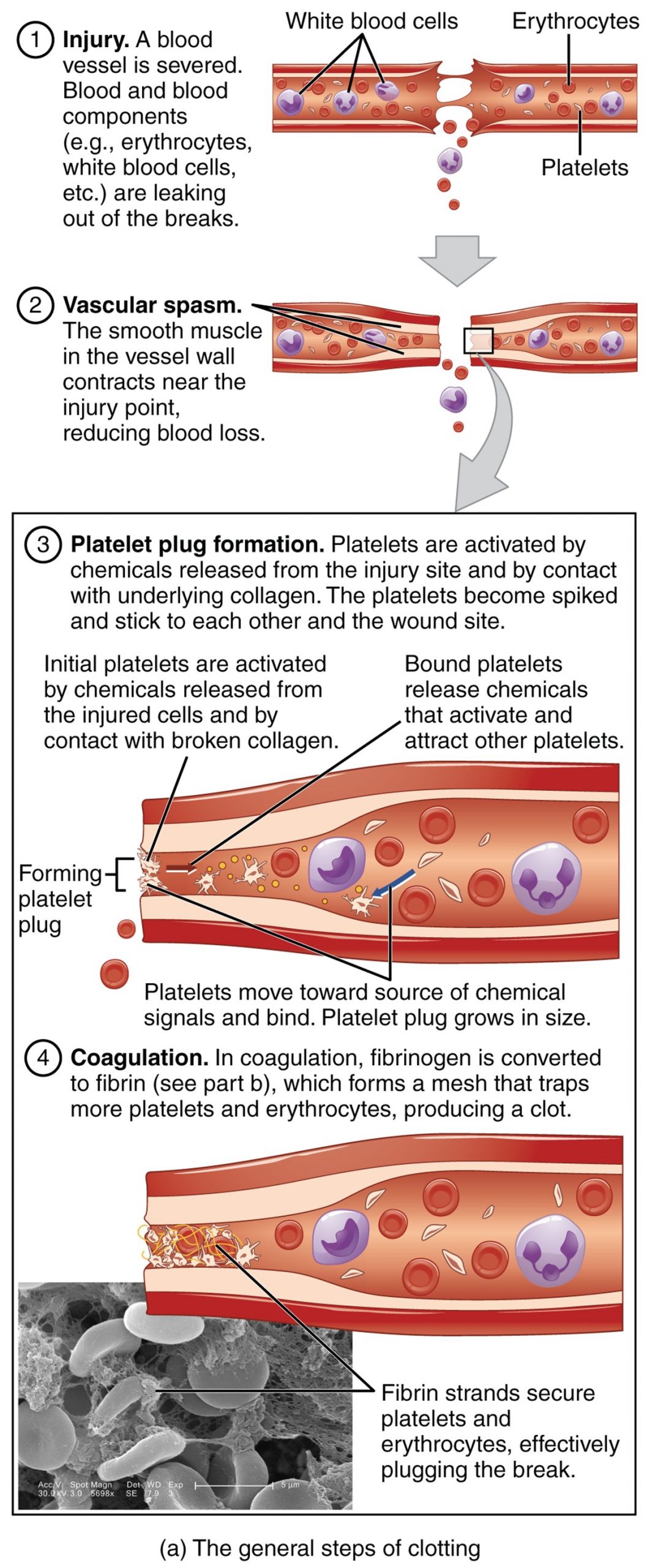
Platelets
After entering the circulation, approximately one-third of the newly-formed platelets migrate to the spleen for storage for later release in response to any rupture in a blood vessel. They then become activated to perform their primary function, which is to limit blood loss. Platelets remain only about 10 days, then are phagocytized by macrophages.
Platelets are key players in hemostasis, the process by which the body seals a ruptured blood vessel and prevents further loss of blood. Although rupture of larger vessels usually requires medical intervention, hemostasis is quite effective in dealing with small, simple wounds. There are three steps to the process: vascular spasm or vasoconstriction, the formation of a platelet plug, and coagulation (blood clotting). Failure of any of these steps will result in hemorrhage. The figure below summarizes the steps of hemostasis.
Figure 7.8 Hemostasis. (a) An injury to a blood vessel initiates the process of hemostasis. Blood clotting involves three steps. First, vascular spasm constricts the flow of blood. Next, a platelet plug forms to temporarily seal small openings in the vessel. Coagulation then enables the repair of the vessel wall once the leakage of blood has stopped. (b) The synthesis of fibrin in blood clots lead to a common pathway. (credit a: Kevin MacKenzie). From Betts, et al., 2021. Licensed under CC BY 4.0.
Fibrinolysis is the process in which a clot is degraded in a healing vessel. An anticoagulant is any substance that opposes coagulation. Several circulating plasma anticoagulants play a role in limiting the coagulation process to the region of injury and restoring a normal, clot-free condition of blood.
Concept Check
Can you explain what happens in each step of hemostasis?
Describe an anticoagulant.
Physiology of Blood
Although carrying oxygen and nutrients to cells and removing wastes from cells is the main function of blood, it is important to realize that blood also serves in defense, distribution of heat, and maintenance of homeostasis.
Transportation
Nutrients from the foods you eat are absorbed in the digestive tract. Most of these travel in the bloodstream directly to the liver, where they are processed and released back into the bloodstream for delivery to body cells.
Oxygen from the air you breathe diffuses into the blood, which moves from the lungs to the heart, which then pumps it out to the rest of the body.
Endocrine glands scattered throughout the body release their products, called hormones, into the bloodstream, which carries them to distant target cells.
Blood also picks up cellular wastes and byproducts, and transports them to various organs for removal. For instance, blood moves carbon dioxide to the lungs for exhalation from the body, and various waste products are transported to the kidneys and liver for excretion from the body in the form of urine or bile.
Defense
Leukocytes protect the organism from disease-causing bacteria, cells with mutated DNA that could multiply to become cancerous, or body cells infected with viruses.
When damage to the vessels results in bleeding, blood platelets and certain proteins dissolved in the plasma, interact to block the ruptured areas of the blood vessels involved. This protects the body from further blood loss.
Homeostasis
If you were exercising on a warm day, your rising core body temperature would trigger several homeostatic mechanisms, including increased transport of blood from your core to your body periphery, which is typically cooler. As blood passes through the vessels of the skin, heat would be dissipated to the environment, and the blood returning to your body core would be cooler. In contrast, on a cold day, blood is diverted away from the skin to maintain a warmer body core. In extreme cases, this may result in frostbite.
Blood helps to regulate the water content of body cells.
Blood also helps to maintain the chemical balance of the body. Proteins and other compounds in blood act as buffers, which thereby help to regulate the pH of body tissues. The pH of blood ranges from 7.35 to 7.45.
Concept Check
These three terms all sound similar. Can you explain them by breaking down the word parts?
Hemostasis
Homeostasis
Hematopoiesis
Blood Vessel Medical Terms Not Easily Broken into Word Parts
Common Cardiovascular System – Blood, Abbreviations
Many terms and phrases related to the cardiovascular system – blood are abbreviated. Learn these common abbreviations by expanding the list below.
Medical Terms in Context
Medical Specialties and Procedures Related to the Blood Vessels and Blood
Vascular Surgeons
Vascular surgery is a specialty in which the physician treats diseases of the blood and lymphatic vessels. This includes repair and replacement of diseased or damaged vessels, removal of plaque from vessels, minimally invasive procedures including the insertion of venous catheters, and traditional surgery (Betts, et al., 2021; Society for Vascular Surgery, n.d.). For more information, please visit Society for Vascular Surgery website.
Hematologists
Hematologists are specialist physicians that diagnose and treat blood disorders. These physicians must be well-versed in a wide array of laboratory procedures, basic medical disciplines, and clinical medicine (American Medical Association, 2019). To learn more about hematologists, visit the American Medical Association’s specialty profile on hematology
Vascular Sonographer
Vascular sonography is a challenging yet rewarding profession. As a sonographer working in this field, you’ll use ultrasound machines to produce images of patients’ veins and arteries using high-frequency sound waves. To learn more, visit the Vascular Sonography Credentials web page.
Phlebotomist
Phlebotomists are professionals trained to draw blood (phleb- = “a blood vessel”; -tomy = “to cut”). When more than a few drops of blood are required, phlebotomists perform a venipuncture, typically of a surface vein in the arm. They perform a capillary stick on a finger, an earlobe, or the heel of an infant when only a small quantity of blood is required. An arterial stick is collected from an artery and used to analyze blood gases. After collection, the blood may be analyzed by medical laboratories or perhaps used for transfusions, donations, or research (Betts, et al., 2021).
Medical Laboratory Scientist/Technician
Medical or clinical laboratories employ a variety of individuals in technical positions. Training is provided through a variety of institutions and certification is through the Canadian Society for Medical Laboratory Science. Specialized positions are:
Medical technologist (MT) tests and analyzes blood, other body fluids, and tissue samples.
Medical laboratory scientists (MLS) perform complex analyses of tissue, blood, and other body fluids.
Medical laboratory assistants (MLA) spend the majority of their time receiving, preparing, testing, and processing specimen samples (American Society for Clinical Pathology, n.d.)
Identify meanings of key word components of the cardiovascular system.
Apply the rules of medical language to pronounce, break into word parts, and define the following terms.
Practice pronouncing and defining these medical terms that are not easily broken into word parts.
Practice pronouncing and defining these commonly abbreviated cardiovascular system terms related to the blood.
Use terms related to the cardiovascular system.
Test your knowledge by answering the questions below.
Chapter Attributions
This chapter was adapted by Karen Hobbs from “Cardiovascular System – Blood Vessels & Blood” by Stacey Grimm; Coleen Allee; Elaine Strachota; Laurie Zielinski; Traci Gotz; Micheal Randolph; and Heidi Belitz. Licensed under a CC BY 4.0 license.
Blood vessels that transport blood away from the heart.
A very small artery that leads to a capillary
A microscopic channel that supplies blood to the tissues through perfusion
The delivery of blood to an area/tissue/organ
Extremely small vein
Blood vessels that carry blood back to the heart.
The smooth muscle layer in the blood vessel wall contracts, causing the vessel diameter to narrow. This increases blood pressure in the vessel.
The smooth muscle layer in the wall of the blood vessel relaxes, allowing the vessel to widen. This decreases blood pressure in the vessel.
large artery in the upper arm near the biceps muscle
Instrument used to measure blood pressure.
The systolic pressure is the higher value (typically around 120 mm Hg) and reflects the arterial pressure resulting from the ejection of blood during ventricular contraction, or systole.
Compliance is the ability of any compartment to expand to accommodate increased content. The greater the compliance of an artery, the more effectively it is able to expand to accommodate surges in blood flow without increased resistance or blood pressure.
Cardiac output is the measurement of blood flow from the heart through the ventricles, and is usually measured in liters per minute. Any factor that causes cardiac output to increase, by elevating heart rate or stroke volume or both, will elevate blood pressure and promote blood flow.
flight or fight response
Viscosity is the thickness of fluids that affects their ability to flow
The number of times the heart contracts in one minute.
A centrifuge is a common piece of laboratory equipment used to spin test tubes at a high speed in order to separate components in a liquid by weight.
red blood cells
also spelled leucocyte, these are white blood cells
also called platelets, these are cell fragments that aid in blood clotting
percentage by volume of red cells in your blood
also "phagocytosed", this is the process by which certain cells are able to 'eat' other cells or substances by engulfing them
a type of leukocyte (usually a monocyte) that has the ability to ingest and destroy other cells or pathogens
excessive or uncontrolled bleeding from the blood vessels
WTCS Learning Objectives
Apply the rules of medical language to build, analyze, spell, pronounce, abbreviate, and define terms as they relate to the nervous system
Identify meanings of key word components of the nervous system
Categorize diagnostic, therapeutic, procedural or anatomic terms related to the nervous system
Use terms related to the nervous system
Use terms related to the diseases and disorders of the nervous system
Nervous System Word Parts
Click on prefixes, combining forms, and suffixes to reveal a list of word parts to memorize for the Nervous System.
Introduction to the Nervous System
The picture you have in your mind of the nervous system probably includes the brain, the nervous tissue contained within the cranium, and the spinal cord, the extension of nervous tissue within the vertebral column. That suggests it is made of two organs—and you may not even think of the spinal cord as an organ—but the nervous system is a very complex structure. Within the brain, many different and separate regions are responsible for many different and separate functions. It is as if the nervous system is composed of many organs that all look similar and can only be differentiated using tools such as the microscope or electrophysiology.
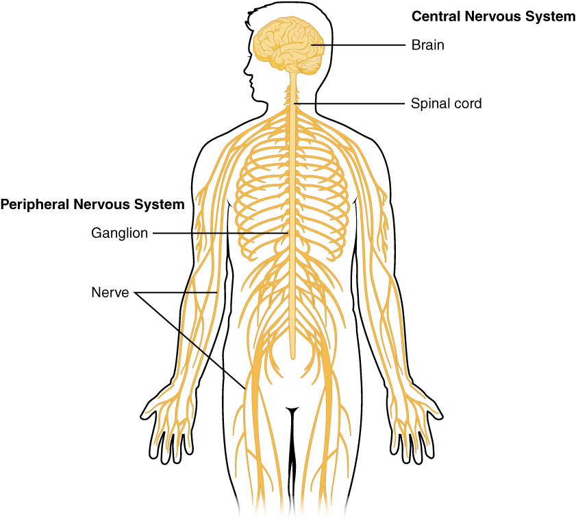
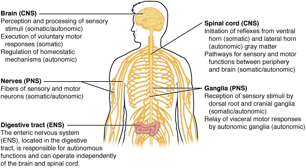
The nervous system can be divided into two major regions: the central and peripheral nervous systems. The central nervous system (CNS) is the brain and spinal cord, and the peripheral nervous system (PNS) is everything else (see Figure 16.1). The brain is contained within the cranial cavity of the skull, and the spinal cord is contained within the vertebral cavity of the vertebral column. It is a bit of an oversimplification to say that the CNS is what is inside these two cavities and the peripheral nervous system is outside of them, but that is one way to start to think about it. In actuality, there are some elements of the peripheral nervous system that are within the cranial or vertebral cavities. The peripheral nervous system is so named because it is on the periphery—meaning beyond the brain and spinal cord. Depending on different aspects of the nervous system, the dividing line between central and peripheral is not necessarily universal.
Figure 16.1 Central and Peripheral Nervous System. The structures of the PNS are referred to as ganglia and nerves, which can be seen as distinct structures. The equivalent structures in the CNS are not obvious from this overall perspective and are best examined in prepared tissue under the microscope. From Betts, et al., 2021. Licensed under CC BY 4.0.
Nervous tissue, present in both the CNS and PNS, contains two basic types of cells: neurons and glial cells. Neurons are the primary type of cell that most anyone associates with the nervous system. They are responsible for the computation and communication that the nervous system provides. They are electrically active and release chemical signals to target cells. Glial cells, or glia, are known to
Did You Know?
The brain has over 100 billion neurons.
play a supporting role for nervous tissue. Ongoing research pursues an expanded role that glial cells might play in signaling, but neurons are still considered the basis of this function. Neurons are important, but without glial support they would not be able to perform their function. A glial cell is one of a variety of cells that provide a framework of tissue that supports the neurons and their activities. The neuron is the more functionally important of the two, in terms of the communicative function of the nervous system. To describe the functional divisions of the nervous system, it is important to understand the structure of a neuron.
Neurons are cells and therefore have a soma, or cell body, but they also have extensions of the cell; each extension is generally referred to as a process. There is one important process that every neuron has called an axon, which is the fiber that connects a neuron with its target. Another type of process that branches off from the soma is the dendrite. Dendrites are responsible for receiving most of the input from other neurons.
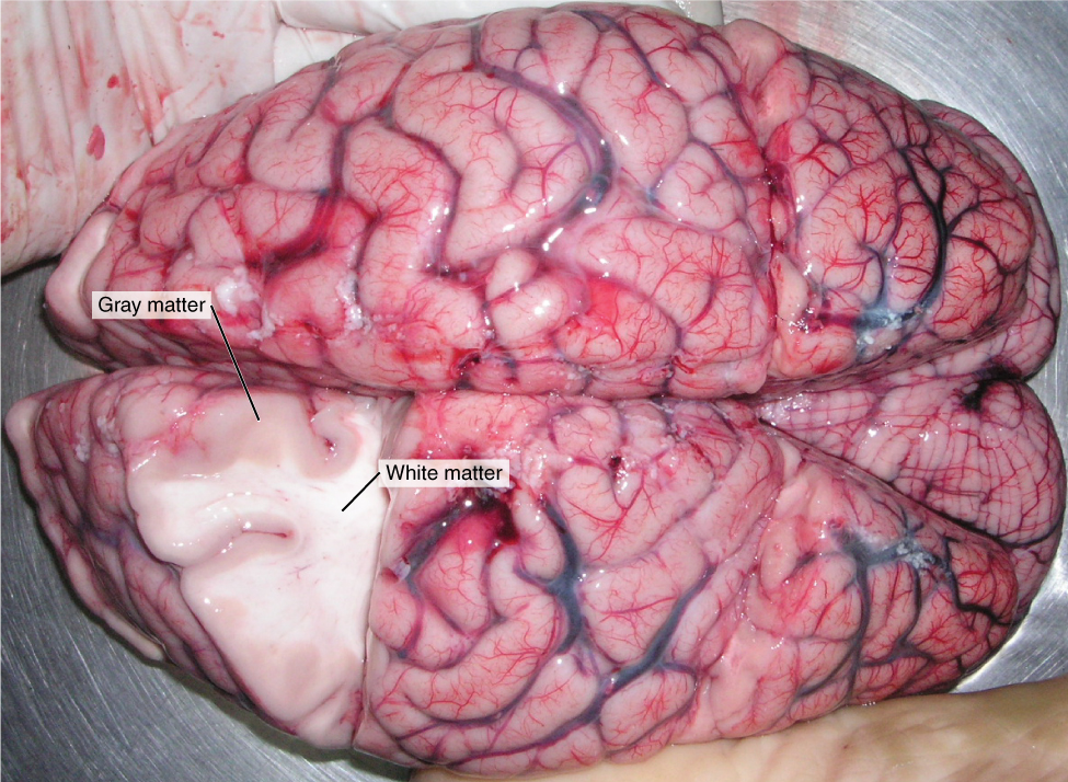
Looking at nervous tissue, there are regions that predominantly contain cell bodies and regions that are largely composed of just axons. These two regions within nervous system structures are often referred to as gray matter (the regions with many cell bodies and dendrites) or white matter (the regions with many axons). Figure 16.2 demonstrates the appearance of these regions in the brain and spinal cord. The colors ascribed to these regions are what would be seen in “fresh,” or unstained, nervous tissue. Gray matter is not necessarily gray. It can be pinkish because of blood content, or even slightly tan, depending on how long the tissue has been preserved. White matter is white because axons are insulated by a lipid-rich substance called myelin. Lipids can appear as white (“fatty”) material, much like the fat on a raw piece of chicken or beef. Actually, gray matter may have that color ascribed to it because next to the white matter, it is just darker—hence, gray.
The distinction between gray matter and white matter is most often applied to central nervous tissue, which has large regions that can be seen with the unaided eye. When looking at peripheral structures, often a microscope is used and the tissue is stained with artificial colors. That is not to say that central nervous tissue cannot be stained and viewed under a microscope, but unstained tissue is most likely from the CNS—for example, a frontal section of the brain or cross section of the spinal cord.
Figure 16.2 Gray Matter and White Matter. A brain removed during an autopsy, with a partial section removed, shows white matter surrounded by gray matter. Gray matter makes up the outer cortex of the brain. (credit: modification of work by “Suseno”/Wikimedia Commons). From Betts, et al., 2021. Licensed under CC BY 4.0.
The Adult Brain
The adult brain is separated into four major regions: the cerebrum, the diencephalon, the brain stem, and the cerebellum. The cerebrum is the largest portion and contains the cerebral cortex and subcortical nuclei. It is divided into two halves by the longitudinal fissure.
The Cerebrum
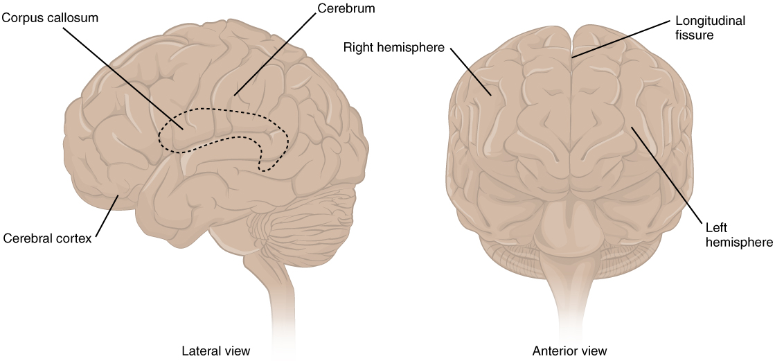
The iconic gray mantle of the human brain, which appears to make up most of the mass of the brain, is the cerebrum (see Figure 16.3). The wrinkled portion is the cerebral cortex, and the rest of the structure is beneath that outer covering. There is a large separation between the two sides of the cerebrum called the longitudinal fissure. It separates the cerebrum into two distinct halves, a right and left cerebral hemisphere. Deep within the cerebrum, the white matter of the corpus callosum provides the major pathway for communication between the two hemispheres of the cerebral cortex.
Figure 16.3 The Cerebrum. The cerebrum is a large component of the CNS in humans, and the most obvious aspect of it is the folded surface called the cerebral cortex. From Betts, et al., 2021. Licensed under CC BY 4.0.
Did You Know?
The brain is about 75% water and is the fattest organ in the body.
Many of the higher neurological functions, such as memory, emotion, and consciousness, are the result of cerebral function. The complexity of the cerebrum is different across vertebrate species. The cerebrum of the most primitive vertebrates is not much more than the connection for the sense of smell. In mammals, the cerebrum comprises the outer gray matter that is the cortex (from the Latin word meaning “bark of a tree”) and several deep nuclei that belong to three important functional groups. The basal nuclei are responsible for cognitive processing, the most important function being that associated with planning movements. The basal forebrain contains nuclei that are important in learning and memory. The limbic cortex is the region of the cerebral cortex that part of the limbic system, a collection of structures involved in emotion, memory, and behavior.
Cerebral Cortex
The cerebrum is covered by a continuous layer of gray matter that wraps around either side of the forebrain—the cerebral cortex. This thin, extensive region of wrinkled gray matter is responsible for the higher functions of the nervous system. A gyrus (plural = gyri) is the ridge of one of those wrinkles, and a sulcus (plural = sulci) is the groove between two gyri. The pattern of these folds of tissue indicates specific regions of the cerebral cortex.
The head is limited by the size of the birth canal, and the brain must fit inside the cranial cavity of the skull. Extensive folding in the cerebral cortex enables more gray matter to fit into this limited space. If the gray matter of the cortex were peeled off of the cerebrum and laid out flat, its surface area would be roughly equal to one square meter.
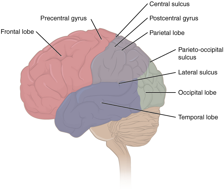
The folding of the cortex maximizes the amount of gray matter in the cranial cavity. During embryonic development, as the telencephalon expands within the skull, the brain goes through a regular course of growth that results in everyone’s brain having a similar pattern of folds. The surface of the brain can be mapped on the basis of the locations of large gyri and sulci. Using these landmarks, the cortex can be separated into four major regions, or lobes (see Figure 16.4). The lateral sulcus that separates the temporal lobe from the other regions is one such landmark. Superior to the lateral sulcus are the parietal lobe and frontal lobe, which are separated from each other by the central sulcus. The posterior region of the cortex is the occipital lobe, which has no obvious anatomical border between it and the parietal or temporal lobes on the lateral surface of the brain. From the medial surface, an obvious landmark separating the parietal and occipital lobes is called the parieto-occipital sulcus. The fact that there is no obvious anatomical border between these lobes is consistent with the functions of these regions being interrelated.
Figure 16.4 Lobes of the Cerebral Cortex. The cerebral cortex is divided into four lobes. Extensive folding increases the surface area available for cerebral functions. From Betts, et al., 2021. Licensed under CC BY 4.0.
Concept Check
Identify the two major divisions of the nervous system.
Describe the cerebral cortex.
What are the halves of the cerebrum know as?
Thalamus
The thalamus is a collection of nuclei that relay information between the cerebral cortex and the periphery, spinal cord, or brain stem. All sensory information, except for the sense of smell, passes through the thalamus before processing by the cortex. For example, the portion of the thalamus that receives visual information will influence what visual stimuli are important, or what receives attention.
The cerebrum also sends information down to the thalamus, which usually communicates motor commands. This involves interactions with the cerebellum and other nuclei in the brain stem. The cerebrum interacts with the basal nuclei, which involves connections with the thalamus. The primary output of the basal nuclei is to the thalamus, which relays that output to the cerebral cortex. The cortex also sends information to the thalamus that will then influence the effects of the basal nuclei.
Hypothalamus
Inferior and slightly anterior to the thalamus is the hypothalamus the other major region of the diencephalon. The hypothalamus is a collection of nuclei that are largely involved in regulating homeostasis. The hypothalamus is the executive region in charge of the autonomic nervous system and the endocrine system through its regulation of the anterior pituitary gland. Other parts of the hypothalamus are involved in memory and emotion as part of the limbic system.
Brain Stem
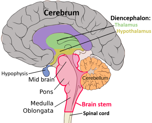
The midbrain and hindbrain (composed of the pons and the medulla) are collectively referred to as the brain stem (see Figure 16.5). The structure emerges from the ventral surface of the forebrain as a tapering cone that connects the brain to the spinal cord. Attached to the brain stem, but considered a separate region of the adult brain, is the cerebellum. The midbrain coordinates sensory representations of the visual, auditory, and somatosensory perceptual spaces. The pons is the main connection with the cerebellum. The pons and the medulla regulate several crucial functions, including the cardiovascular and respiratory systems and rates.
The cranial nerves connect through the brain stem and provide the brain with the sensory input and motor output associated with the head and neck, including most of the special senses. The major ascending and descending pathways between the spinal cord and brain, specifically the cerebrum, pass through the brain stem.
Figure 16.5 Parts of the Brain. The image highlights and names the most important structures of the brain in a sagittal(longitudinal) cross-section. Belomaad Biology Matura book, CC BY-SA 4.0, via Wikimedia Commons
Midbrain
The midbrain is the uppermost portion of the brainstem. It contains pathways connecting the cerebrum with lower portions of the brain and structures involved with seeing and hearing.
Pons
The pons is a part of the brainstem that literally means bridge. It contains nerve fiber tracts that connect the cerebellum and cerebrum with the rest of the brain. Nerves affecting the face and eye movement are located here.
Medulla
The medulla oblongata, also in the brainstem, connects the spinal cord with the rest of the brain. It is the region known as the myelencephalon in the embryonic brain. It contains centers that control respiration, heart rate, and the muscles of the blood vessel walls, which assist in determining blood pressure. Nerve tracts cross from right to left and left to right in the medulla oblongata. For example, nerve cells that control movement of the left side of the body are found in the right half of the cerebrum. These cells send out axons that cross over (decussate) to the opposite side of the brain in the medulla oblongata and then travel down the spinal cord.
The Cerebellum
The cerebellum functions to coordinate voluntary movements and to maintain balance and posture. It is covered in gyri and sulci like the cerebrum, and looks like a miniature version of that part of the brain (see Figure 16.6). The cerebellum is located under the posterior portion of the cerebrum (also called hindbrain). It accounts for approximately 10 percent of the mass of the brain.
Figure 16.6 The Cerebellum. The cerebellum is situated on the posterior surface of the brain stem. Image by Patrick J. Lynch; CC BY 2.5, via Wikimedia Commons
Concept Check
What is the primary processing purpose of the medulla?
Identify the structure in the brain responsible for sensory feedback through the spinal cord. Suggest what may happen if this function failed.
The Spinal Cord
The description of the CNS is concentrated on the structures of the brain, but the spinal cord is another major organ of the system. Whereas the brain develops out of expansions of the neural tube into primary and then secondary vesicles, the spinal cord maintains the tube structure and is only specialized into certain regions. As the spinal cord continues to develop in the newborn, anatomical
Did You Know?
The bundle of nerve fibers making up the spinal cord is no thicker than the human thumb.
features mark its surface. The anterior midline is marked by the anterior median fissure, and the posterior midline is marked by the posterior median sulcus. Axons enter the posterior side through the dorsal (posterior) nerve root, which marks the posterolateral sulcus on either side. The axons emerging from the anterior side do so through the ventral (anterior) nerve root. Note that it is common to see the terms dorsal (dorsal = “back”) and ventral (ventral = “belly”) used interchangeably with posterior and anterior, particularly in reference to nerves and the structures of the spinal cord. You should learn to be comfortable with both.
On the whole, the posterior regions are responsible for sensory functions and the anterior regions are associated with motor functions. This comes from the initial development of the spinal cord, which is divided into the basal plate and the alar plate. The basal plate is closest to the ventral midline of the neural tube, which will become the anterior face of the spinal cord and gives rise to motor neurons. The alar plate is on the dorsal side of the neural tube and gives rise to neurons that will receive sensory input from the periphery.
The length of the spinal cord is divided into regions that correspond to the regions of the vertebral column. The name of a spinal cord region corresponds to the level at which spinal nerves pass through the intervertebral foramina. Immediately adjacent to the brain stem is the following divisions of the spinal cord:
cervical region
thoracic region
lumbar region
sacral region
The spinal cord is not the full length of the vertebral column because the spinal cord does not grow significantly longer after the first or second year, but the skeleton continues to grow. The nerves that emerge from the spinal cord pass through the intervertebral formina at the respective levels. As the vertebral column grows, these nerves grow with it and result in a long bundle of nerves that resembles a horse’s tail and is named the cauda equina. The sacral spinal cord is at the level of the upper lumbar vertebral bones. The spinal nerves extend from their various levels to the proper level of the vertebral column.
Neurons
Neurons are the cells considered to be the basis of nervous tissue. They are responsible for the electrical signals that communicate information about sensations, and that produce movements in response to those stimuli, along with inducing thought processes within the brain. An important part of the function of neurons is in their structure, or shape. The three-dimensional shape of these cells makes the immense numbers of connections within the nervous system possible.
Parts of a Neuron
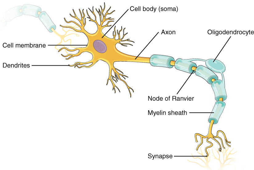
As you learned in the first section, the main part of a neuron is the cell body, which is also known as the soma (soma = “body”). The cell body contains the nucleus and most of the major organelles. But what makes neurons special is that they have many extensions of their cell membranes, which are generally referred to as processes. Neurons are usually described as having one, and only one, axon—a fiber that emerges from the cell body and projects to target cells. That single axon can branch repeatedly to communicate with many target cells. It is the axon that propagates the nerve impulse, which is communicated to one or more cells. The other processes of the neuron are dendrites, which receive information from other neurons at specialized areas of contact called synapses. The dendrites are usually highly branched processes, providing locations for other neurons to communicate with the cell body. Information flows through a neuron from the dendrites, across the cell body, and down the axon. This gives the neuron a polarity—meaning that information flows in this one direction. Figure 16.7 shows the relationship of these parts to one another.
Figure 16.7 Parts of a Neuron. The major parts of the neuron are labeled on a multipolar neuron from the CNS. From Betts, et al., 2021. Licensed under CC BY 4.0.
Many axons are wrapped by an insulating substance called myelin, which is actually made from glial cells. Myelin acts as insulation much like the plastic or rubber that is used to insulate electrical wires. A key difference between myelin and the insulation on a wire is that there are gaps in the myelin covering of an axon. Each gap is called a node of Ranvier and is important to the way that electrical signals travel down the axon. The length of the axon between each gap, which is wrapped in myelin, is referred to as an axon segment. At the end of the axon is the axon terminal, where there are usually several branches extending toward the target cell, each of which ends in an enlargement called a synaptic end bulb. These bulbs are what make the connection with the target cell at the synapse.
Types of Neurons
There are many neurons in the nervous system—a number in the trillions. And there are many different types of neurons. They can be classified by many different criteria. The first way to classify them is by the number of processes attached to the cell body. Using the standard model of neurons, one of these processes is the axon, and the rest are dendrites. Because information flows through the neuron from dendrites or cell bodies toward the axon, these names are based on the neuron's polarity.
Glial Cells
Glial cells, or neuroglia or simply glia, are the other type of cell found in nervous tissue. They are considered to be supporting cells, and many functions are directed at helping neurons complete their function for communication. The name glia comes from the Greek word that means “glue,” and was coined by the German pathologist Rudolph Virchow, who wrote in 1856: “This connective substance, which is in the brain, the spinal cord, and the special sense nerves, is a kind of glue (neuroglia) in which the nervous elements are planted.” Today, research into nervous tissue has shown that there are many deeper roles that these cells play. And research may find much more about them in the future.
There are six types of glial cells. Four of them are found in the CNS and two are found in the PNS. Table 16.1 outlines some common characteristics and functions.
Table 16.1: Glial Cell Types by Location and Basic Function. From Betts, et al., 2021. Licensed under CC BY 4.0.
CNS GLIA
PNS GLIA
BASIC FUNCTION
Astrocyte
Satellite cell
Support
Oligodendrocyte
Schwann cell
Insulation, myelination
Microglia
-
Immune surveillance and phagocytosis
Ependymal cell
-
Creating CSF
Glial Cells of the CNS
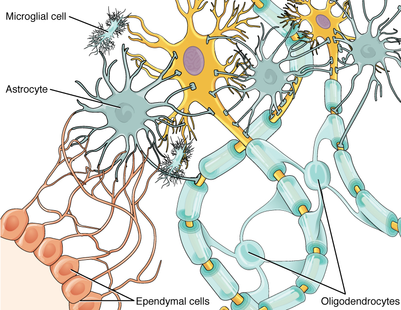
One cell providing support to neurons of the CNS is the astrocyte, so named because it appears to be star-shaped under the microscope (astro- = “star”). Astrocytes have many processes extending from their main cell body (not axons or dendrites like neurons, just cell extensions). Those processes extend to interact with neurons, blood vessels, or the connective tissue covering the CNS that is called the pia mater (see Figure 16.8). Generally, they are supporting cells for the neurons in the central nervous system. Some ways in which they support neurons in the central nervous system are by maintaining the concentration of chemicals in the extracellular space, removing excess signaling molecules, reacting to tissue damage, and contributing to the blood-brain barrier (BBB). The blood-brain barrier is a physiological barrier that keeps many substances that circulate in the rest of the body from getting into the central nervous system, restricting what can cross from circulating blood into the CNS. Nutrient molecules, such as glucose or amino acids, can pass through the BBB, but other molecules cannot. This actually causes problems with drug delivery to the CNS. Pharmaceutical companies are challenged to design drugs that can cross the BBB as well as have an effect on the nervous system.
Figure 16.8 Glial Cells of the CNS. The CNS has astrocytes, oligodendrocytes, microglia, and ependymal cells that support the neurons of the CNS in several ways. From Betts, et al., 2021. Licensed under CC BY 4.0.
Like a few other parts of the body, the brain has a privileged blood supply. Very little can pass through by diffusion. Most substances that cross the wall of a blood vessel into the CNS must do so through an active transport process. Because of this, only specific types of molecules can enter the CNS. Glucose—the primary energy source—is allowed, as are amino acids. Water and some other small particles, like gases and ions, can enter. But most everything else cannot, including white blood cells, which are one of the body’s main lines of defense. While this barrier protects the CNS from exposure to toxic or pathogenic substances, it also keeps out the cells that could protect the brain and spinal cord from disease and damage. The BBB also makes it harder for pharmaceuticals to be developed that can affect the nervous system. Aside from finding efficacious substances, the means of delivery is also crucial.
Oligodendrocyte, sometimes called just “oligo,” which is the glial cell type that insulates axons in the CNS. The name means “cell of a few branches” (oligo- = “few”; dendro- = “branches”; -cyte = “cell”).
Microglia are smaller than most of the other glial cells. Ongoing research into these cells, although not entirely conclusive, suggests that they may originate as white blood cells, called macrophages, that become part of the CNS during early development. Their function is related to what macrophages do in the rest of the body. When macrophages encounter diseased or damaged cells in the rest of the body, they ingest and digest those cells or the pathogens that cause disease. Microglia are the cells in the CNS that can do this in normal, healthy tissue, and they are therefore also referred to as CNS-resident macrophages.
The ependymal cell is a glial cell that filters blood to make cerebrospinal fluid (CSF), the fluid that circulates through the CNS. Because of the privileged blood supply inherent in the BBB, the extracellular space in nervous tissue does not easily exchange components with the blood. Ependymal cells line each ventricle, one of four central cavities that are remnants of the hollow center of the neural tube formed during the embryonic development of the brain. They also have cilia on their apical surface to help move the CSF through the ventricular space. The relationship of these glial cells to the structure of the CNS is seen in Figure 16.8.
Glial Cells of the PNS
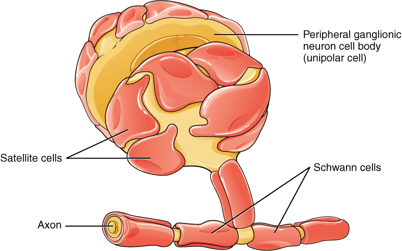
One of the two types of glial cells found in the PNS is the satellite cell. Satellite cells are found in sensory and autonomic ganglia, where they surround the cell bodies of neurons. This accounts for the name, based on their appearance under the microscope. They provide support, performing similar functions in the periphery as astrocytes do in the CNS—except, of course, for establishing the BBB.
The second type of glial cell is the Schwann cell, which insulate axons with myelin in the periphery. Schwann cells are different than oligodendrocytes, in that a Schwann cell wraps around a portion of only one axon segment and no others. Oligodendrocytes have processes that reach out to multiple axon segments, whereas the entire Schwann cell surrounds just one axon segment. The nucleus and cytoplasm of the Schwann cell are on the edge of the myelin sheath. The relationship of these two types of glial cells to ganglia and nerves in the PNS is seen in Figure 16.9.
Figure 16.9 Glial Cells of the PNS. The PNS has satellite cells and Schwann cells. From Betts, et al., 2021. Licensed under CC BY 4.0.
Myelin
The appearance of the myelin sheath can be thought of as similar to the pastry wrapped around a hot dog for “pigs in a blanket” or a similar food. The glial cell is wrapped around the axon several times with little to no cytoplasm between the glial cell layers. For oligodendrocytes, the rest of the cell is separate from the myelin sheath as a cell process extends back toward the cell body. A few other processes provide the same insulation for other axon segments in the area. For Schwann cells, the outermost layer of the cell membrane contains cytoplasm and the nucleus of the cell as a bulge on one side of the myelin sheath. During development, the glial cell is loosely or incompletely wrapped around the axon. The edges of this loose enclosure extend toward each other, and one end tucks under the other. The inner edge wraps around the axon, creating several layers, and the other edge closes around the outside so that the axon is completely enclosed.
Anatomy Labeling Activity
Physiology (Function) of the Nervous System
The nervous system is involved in receiving information about the environment around us (sensation) and generating responses to that information (motor responses). The nervous system can be divided into regions that are responsible for sensation (sensory functions) and for the response (motor functions). But there is a third function that needs to be included. Sensory input needs to be integrated with other sensations, as well as with memories, emotional state, or learning (cognition). Some regions of the nervous system are termed integration or association areas. The process of integration combines sensory perceptions and higher cognitive functions such as memories, learning, and emotion to produce a response.
Sensation
The first major function of the nervous system is sensation—receiving information about the environment to gain input about what is happening outside the body (or, sometimes, within the body). The sensory functions of the nervous system register the presence of a change from homeostasis or a particular event in the environment, known as a stimulus. The senses we think of most are the “big five”: taste, smell, touch, sight, and hearing. The stimuli for taste and smell are both chemical substances (molecules, compounds, ions, etc.), touch is physical or mechanical stimuli that interact with the skin, sight is light stimuli, and hearing is the perception of sound, which is a physical stimulus similar to some aspects of touch. There are actually more senses than just those, but that list represents the major senses. Those five are all senses that receive stimuli from the outside world, and of which there is conscious perception. Additional sensory stimuli might be from the internal environment (inside the body), such as the stretch of an organ wall or the concentration of certain ions in the blood.
Response
The nervous system produces a response on the basis of the stimuli perceived by sensory structures. An obvious response would be the movement of muscles, such as withdrawing a hand from a hot stove, but there are broader uses of the term. The nervous system can cause the contraction of all three types of muscle tissue. For example, skeletal muscle contracts to move the skeleton, cardiac muscle is influenced as heart rate increases during exercise, and smooth muscle contracts as the digestive system moves food along the digestive tract. Responses also include the neural control of glands in the body as well, such as the production and secretion of sweat by the sweat glands found in the skin to lower body temperature.
Responses can be divided into those that are voluntary or conscious (contraction of skeletal muscle) and those that are involuntary (contraction of smooth muscles, regulation of cardiac muscle, activation of glands). Voluntary responses are governed by the somatic nervous system and involuntary responses are governed by the autonomic nervous system, which are discussed in the next section.
Integration
Stimuli that are received by sensory structures are communicated to the nervous system where that information is processed. This is called integration. Stimuli are compared with, or integrated with, other stimuli, memories of previous stimuli, or the state of a person at a particular time. This leads to the specific response that will be generated. Seeing a baseball pitched to a batter will not automatically cause the batter to swing. The trajectory of the ball and its speed will need to be considered. Maybe the count is three balls and one strike, and the batter wants to let this pitch go by in the hope of getting a walk to first base. Or maybe the batter’s team is so far ahead, it would be fun to just swing away.
Controlling the Body
The nervous system can be divided into two parts mostly on the basis of a functional difference in responses. The somatic nervous system (SNS) is responsible for conscious perception and voluntary motor responses. Voluntary motor response means the contraction of skeletal muscle, but those contractions are not always voluntary in the sense that you have to want to perform them. Some somatic motor responses are reflexes, and often happen without a conscious decision to perform them. If your friend jumps out from behind a corner and yells “Boo!” you will be startled and you might scream or leap back. You didn’t decide to do that, and you may not have wanted to give your friend a reason to laugh at your expense, but it is a reflex involving skeletal muscle contractions. Other motor responses become automatic (in other words, unconscious) as a person learns motor skills (referred to as “habit learning” or “procedural memory”).
The autonomic nervous system (ANS) is responsible for involuntary control of the body, usually for the sake of homeostasis (regulation of the internal environment). Sensory input for autonomic functions can be from sensory structures tuned to external or internal environmental stimuli. The motor output extends to smooth and cardiac muscle as well as glandular tissue. The role of the autonomic system is to regulate the organ systems of the body, which usually means to control homeostasis. Sweat glands, for example, are controlled by the autonomic system. When you are hot, sweating helps cool your body down. That is a homeostatic mechanism. But when you are nervous, you might start sweating also. That is not homeostatic, it is the physiological response to an emotional state.
There is another division of the nervous system that describes functional responses. The enteric nervous system (ENS) is responsible for controlling the smooth muscle and glandular tissue in your digestive system. It is a large part of the PNS, and is not dependent on the CNS. It is sometimes valid, however, to consider the enteric system to be a part of the autonomic system because the neural structures that make up the enteric system are a component of the autonomic output that regulates digestion. There are some differences between the two, but for our purposes here there will be a good bit of overlap. See Figure 16.10 for examples of where these divisions of the nervous system can be found.
Figure 16.10 Somatic, Autonomic, and Enteric Structures of the Nervous System. Somatic structures include the spinal nerves, both motor and sensory fibers, as well as the sensory ganglia (posterior root ganglia and cranial nerve ganglia). Autonomic structures are found in the nerves also, but include the sympathetic and parasympathetic ganglia. The enteric nervous system includes the nervous tissue within the organs of the digestive tract. From Betts, et al., 2021. Licensed under CC BY 4.0.
Functions of the Cerebral Cortex
The cerebrum is the seat of many of the higher mental functions, such as memory and learning, language, and conscious perception, which are the subjects of subtests of the mental status exam. The cerebral cortex is the thin layer of gray matter on the outside of the cerebrum. It is approximately a millimeter thick in most regions and highly folded to fit within the limited space of the cranial vault. These higher functions are distributed across various regions of the cortex, and specific locations can be said to be responsible for particular functions. There is a limited set of regions, for example, that are involved in language function, and they can be subdivided on the basis of the particular part of language function that each governs.
Cognitive Abilities
Assessment of cerebral functions is directed at cognitive abilities. The abilities assessed through the mental status exam can be separated into four groups: orientation and memory, language and speech, sensorium, and judgment and abstract reasoning.
Orientation and Memory
Orientation is the patient’s awareness of his or her immediate circumstances. It is awareness of time, not in terms of the clock, but of the date and what is occurring around the patient. It is awareness of place, such that a patient should know where he or she is and why. It is also awareness of who the patient is—recognizing personal identity and being able to relate that to the examiner. The initial tests of orientation are based on the questions, “Do you know what the date is?” or “Do you know where you are?” or “What is your name?” Further understanding of a patient’s awareness of orientation can come from questions that address remote memory, such as “Who is the President of the United States?”, or asking what happened on a specific date.
Memory is largely a function of the temporal lobe, along with structures beneath the cerebral cortex such as the hippocampus and the amygdala. The storage of memory requires these structures of the medial temporal lobe. A famous case of a man who had both medial temporal lobes removed to treat intractable epilepsy provided insight into the relationship between the structures of the brain and the function of memory.
The prefrontal cortex can also be tested for the ability to organize information. In one subtest of the mental status exam called set generation, the patient is asked to generate a list of words that all start with the same letter, but not to include proper nouns or names. The expectation is that a person can generate such a list of at least 10 words within 1 minute. Many people can likely do this much more quickly, but the standard separates the accepted normal from those with compromised prefrontal cortices.
Read this article to learn about a young man who texts his fiancée in a panic as he finds that he is having trouble remembering things. At the hospital, a neurologist administers the mental status exam, which is mostly normal except for the three-word recall test. The young man could not recall them even 30 seconds after hearing them and repeating them back to the doctor. An undiscovered mass in the mediastinum region was found to be Hodgkin’s lymphoma, a type of cancer that affects the immune system and likely caused antibodies to attack the nervous system. The patient eventually regained his ability to remember, though the events in the hospital were always elusive. Considering that the effects on memory were temporary, but resulted in the loss of the specific events of the hospital stay, what regions of the brain were likely to have been affected by the antibodies and what type of memory does that represent?
Language and Speech
Language is, arguably, a very human aspect of neurological function. There are certainly strides being made in understanding communication in other species, but much of what makes the human experience seemingly unique is its basis in language. Any understanding of our species is necessarily reflective, as suggested by the question “What am I?” And the fundamental answer to this question is suggested by the famous quote by René Descartes: “Cogito Ergo Sum” (translated from Latin as “I think, therefore I am”). Formulating an understanding of yourself is largely describing who you are to yourself. It is a confusing topic to delve into, but language is certainly at the core of what it means to be self-aware.
The neurological exam has two specific subtests that address language. One measures the ability of the patient to understand language by asking them to follow a set of instructions to perform an action, such as “touch your right finger to your left elbow and then to your right knee.” Another subtest assesses the fluency and coherency of language by having the patient generate descriptions of objects or scenes depicted in drawings, and by reciting sentences or explaining a written passage.
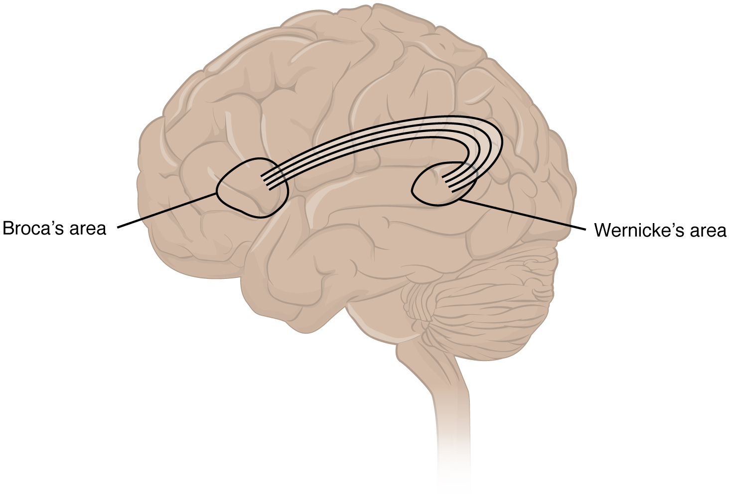
An important example of multimodal integrative areas is associated with language function (see Figure 16.11). Adjacent to the auditory association cortex, at the end of the lateral sulcus just anterior to the visual cortex, is Wernicke’s area. In the lateral aspect of the frontal lobe, just anterior to the region of the motor cortex associated with the head and neck, is Broca’s area. Both regions were originally described on the basis of losses of speech and language, which is called aphasia. The aphasia associated with Broca’s area is known as an expressive aphasia, which means that speech production is compromised. This type of aphasia is often described as non-fluency because the ability to say some words leads to broken or halting speech. Grammar can also appear to be lost. The aphasia associated with Wernicke’s area is known as a receptive aphasia, which is not a loss of speech production, but a loss of understanding of content. Patients, after recovering from acute forms of this aphasia, report not being able to understand what is said to them or what they are saying themselves, but they often cannot keep from talking.
The two regions are connected by white matter tracts that run between the posterior temporal lobe and the lateral aspect of the frontal lobe. Conduction aphasia associated with damage to this connection refers to the problem of connecting the understanding of language to the production of speech. This is a very rare condition, but is likely to present as an inability to faithfully repeat spoken language.
Figure 16.11 Broca's and Wernicke's Areas. Two important integration areas of the cerebral cortex associated with language function are Broca’s and Wernicke’s areas. The two areas are connected through the deep white matter running from the posterior temporal lobe to the frontal lobe. From Betts, et al., 2021. Licensed under CC BY 4.0.
Sensorium
Those parts of the brain involved in the reception and interpretation of sensory stimuli are referred to collectively as the sensorium. The cerebral cortex has several regions that are necessary for sensory perception. Several of the subtests can reveal activity associated with these sensory modalities, such as being able to hear a question or see a picture. Two subtests assess specific functions of these cortical areas.
The first is praxis, a practical exercise in which the patient performs a task completely on the basis of verbal description without any demonstration from the examiner. The second subtest for sensory perception is gnosis, which involves two tasks. The first task, known as stereognosis, involves the naming of objects strictly on the basis of the somatosensory information that comes from manipulating them. The patient keeps their eyes closed and is given a common object, such as a coin, that they have to identify. The patient should be able to indicate the particular type of coin, such as a dime versus a penny, or a nickel versus a quarter, on the basis of the sensory cues involved. For example, the size, thickness, or weight of the coin may be an indication, or to differentiate the pairs of coins suggested here, the smooth or corrugated edge of the coin will correspond to the particular denomination. The second task, graphesthesia, is to recognize numbers or letters written on the palm of the hand with a dull pointer, such as a pen cap.
Judgment and Abstract Reasoning
Planning and producing responses requires an ability to make sense of the world around us. Making judgments and reasoning in the abstract are necessary to produce movements as part of larger responses. For example, when your alarm goes off, do you hit the snooze button or jump out of bed? Is 10 extra minutes in bed worth the extra rush to get ready for your day? Will hitting the snooze button multiple times lead to feeling more rested or result in a panic as you run late? How you mentally process these questions can affect your whole day.
The prefrontal cortex is responsible for the functions responsible for planning and making decisions. In the mental status exam, the subtest that assesses judgment and reasoning is directed at three aspects of frontal lobe function. First, the examiner asks questions about problem solving, such as “If you see a house on fire, what would you do?” The patient is also asked to interpret common proverbs, such as “Don’t look a gift horse in the mouth.” Additionally, pairs of words are compared for similarities, such as apple and orange, or lamp and cabinet.
Everyday Connections
Left Brain, Right Brain
Popular media often refer to right-brained and left-brained people, as if the brain were two independent halves that work differently for different people. This is a popular misinterpretation of an important neurological phenomenon. As an extreme measure to deal with a debilitating condition, the corpus callosum may be sectioned to overcome intractable epilepsy. When the connections between the two cerebral hemispheres are cut, interesting effects can be observed.
The reason for this is that the language functions of the cerebral cortex are localized to the left hemisphere in 95 percent of the population. Additionally, the left hemisphere is connected to the right side of the body through the corticospinal tract and the ascending tracts of the spinal cord. Motor commands from the precentral gyrus control the opposite side of the body, whereas sensory information processed by the postcentral gyrus is received from the opposite side of the body. For a verbal command to initiate movement of the right arm and hand, the left side of the brain needs to be connected by the corpus callosum. Language is processed in the left side of the brain and directly influences the left brain and right arm motor functions, but is sent to influence the right brain and left arm motor functions through the corpus callosum. Likewise, the left-handed sensory perception of what is in the left pocket travels across the corpus callosum from the right brain, so no verbal report on those contents would be possible if the hand happened to be in the pocket.
People who have had their corpus callosum cut can perform two independent tasks at the same time because the lines of communication between the right and left sides of his brain have been removed. Whereas a person with an intact corpus callosum cannot overcome the dominance of one hemisphere over the other, this patient can. If the left cerebral hemisphere is dominant in the majority of people, why would right-handedness be most common?
A class of disorders that affect the nervous system are the neurodegenerative diseases: Alzheimer’s disease, Parkinson’s disease, Huntington’s disease, amyotrophic lateral sclerosis (ALS), Creutzfeld–Jacob disease, multiple sclerosis (MS), and other disorders that are the result of nervous tissue degeneration. In diseases like Alzheimer’s, Parkinson’s, or ALS, neurons die; in diseases like MS, myelin is affected. Some of these disorders affect motor function, and others present with dementia. Some are the result of genetics, such as Huntington’s disease, or the result of autoimmunity, such as MS; others are not entirely understood, such as Alzheimer’s and Parkinson’s diseases.
Several diseases can result from the demyelination of axons. The causes of these diseases are not the same; some have genetic causes, some are caused by pathogens, and others are the result of autoimmune disorders. Though the causes are varied, the results are largely similar. The myelin insulation of axons is compromised, making electrical signaling slower (Betts, et al., 2021).
Multiple sclerosis (MS) is one such disease. It is an example of an autoimmune disease. The antibodies produced by lymphocytes (a type of white blood cell) mark myelin as something that should not be in the body. This causes inflammation and the destruction of the myelin in the central nervous system. As the insulation around the axons is destroyed by the disease, scarring becomes obvious (Betts, et al., 2021).
Guillain-Barre (pronounced gee-YAN bah-RAY) syndrome is an example of a demyelinating disease of the peripheral nervous system. It is also the result of an autoimmune reaction, but the inflammation is in peripheral nerves. Sensory symptoms or motor deficits are common, and autonomic failures can lead to changes in the heart rhythm or a drop in blood pressure, especially when standing, which causes dizziness (Betts, et al., 2021).
Other Nerve Disorders
Infection, trauma, and congenital disorders can all lead to significant signs, as identified through the neurological exam. It is important to differentiate between an acute event, such as stroke, and a chronic or global condition such as blunt force trauma. Responses seen in the neurological exam can help. A loss of language function observed in all its aspects is more likely a global event as opposed to a discrete loss of one function, such as not being able to say certain types of words. A concern, however, is that a specific function—such as controlling the muscles of speech—may mask other language functions. The various subtests within the mental status exam can address these finer points and help clarify the underlying cause of the neurological loss (Betts, et al., 2021).
Stroke
Damage to the nervous system can be limited to individual structures or can be distributed across broad areas of the brain and spinal cord. Localized, limited injury to the nervous system is most often the result of circulatory problems. The loss of blood flow to part of the brain is known as a stroke, or a cerebrovascular accident (CVA). There are two main types of stroke, depending on how the blood supply is compromised: ischemic and hemorrhagic. An ischemic stroke is the loss of blood flow to an area because vessels are blocked or narrowed. This is often caused by an embolus, which may be a blood clot or fat deposit. Ischemia may also be the result of thickening of the blood vessel wall, or a drop in blood volume in the brain known as hypovolemia. A hemorrhagic stroke is bleeding into the brain because of a damaged blood vessel. Accumulated blood fills a region of the cranial vault and presses against the tissue in the brain (see Figure 16.12) (Betts, et al., 2021).
Figure 16.12 Hemorrhagic Stroke. (a) A hemorrhage into the tissue of the cerebrum results in a large accumulation of blood with an additional edema in the adjacent tissue. The hemorrhagic area causes the entire brain to be disfigured as suggested here by the lateral ventricles being squeezed into the opposite hemisphere. (b) A CT scan shows an intraparenchymal hemorrhage within the parietal lobe. (credit b: James Heilman). From Betts, et al., 2021. Licensed under CC BY 4.0.
Cerebral Palsy
Cerebral Palsy (CP) is caused by an interruption to the normal development of a person's brain leading to weakness with muscles. Depending on the area of the brain that is affected, signs and symptoms will vary in the type and severity between individuals. Balance and coordination are often challenging due the inability to control muscles (Centers for Disease Control and Prevention, 2019). To learn more about cerebral palsy please visit the Centers for Disease Control and Prevention's web page on cerebral palsy.
Traumatic Brain Injury (TBI)
According to the Centers for Disease Control and Prevention, approximately 61,000 Americans die each year due to causes related to traumatic brain injuries. Brain injuries range from moderate to severe and include concussions. TBI can be caused by falls, automobile accidents, sports, assaults and strokes. Investment has been made to educate people on how to prevent TBIs with a focus on concussions from sports (Centers for Disease Control and Prevention, 2021).
Medical Terms in Context
Medical Specialties
Primary Specialist - Neurologist
Neurologists are medical doctors who complete an additional specialized training in the prevention, diagnosis, and treatment of disorders and conditions related to the brain, spinal cord, nerves and muscles (American Academy of Neurology, 2021). For more details visit the American Academy of Neurology’s webpage.
Procedures Related to the Nervous System
Lumbar Puncture (Spinal Tap)
A neurologist may order this procedure to test cerebrospinal fluid (CSF). This procedure is recommended if they believe symptoms are caused by a problem in the nervous system that can be detected in the cerebrospinal fluid. The procedure involves inserting a needle into the spine after numbing it and taking a sample of cerebrospinal fluid (Mayo Clinic, 2020).
Tensilon Test
This procedure can help a neurologist diagnose myasthenia gravis. In this test, the doctor injects with a medicine called Tensilon. Then they observe how it affects muscle movements (Bergen, 2018). For more information visit Healthline's Tensilon Test web page.
Electromyography (EMG)
An EMG measures electrical activity between your brain or spinal cord to a peripheral nerve. This nerve is found in your arms and legs, and is responsible for muscle control during times of movement and rest. EMGs can help your neurologist diagnose spinal cord disease as well as general muscle or nerve dysfunction (Moores & Cirino, 2018).
Electroencephalogram (EEG)
With electrodes applied to your scalp, an EEG measures electrical activity in the brain. It’s used to help diagnose conditions of the brain, including inflammation, tumors, and injuries, as well as seizures and psychiatric disorders.
Test Yourself
The original version of this chapter contained H5P content. You may want to remove or replace this element.