
VIRTUAL GRAM STAIN LAB EXERCISE
LEARNING OBJECTIVES
Properly perform the Gram staining technique.
Recognize morphology of bacteria.
Differentiate Gram-positive and Gram-negative cell envelopes.
Explain the importance of Gram stains in a clinical environment.
Explain the function of each reagent used in a Gram stain and its correlation with cell envelope structure.
MCCCD OFFICIAL COURSE COMPETENCIES
Describe the structural characteristics of the major groups of microorganisms.
PHOTO REQUIREMENTS
Take a photo with your photo ID during the lab exercises when you see this icon.

Paste the photos in the Virtual Gram Stain Questions Document.
the gram stain
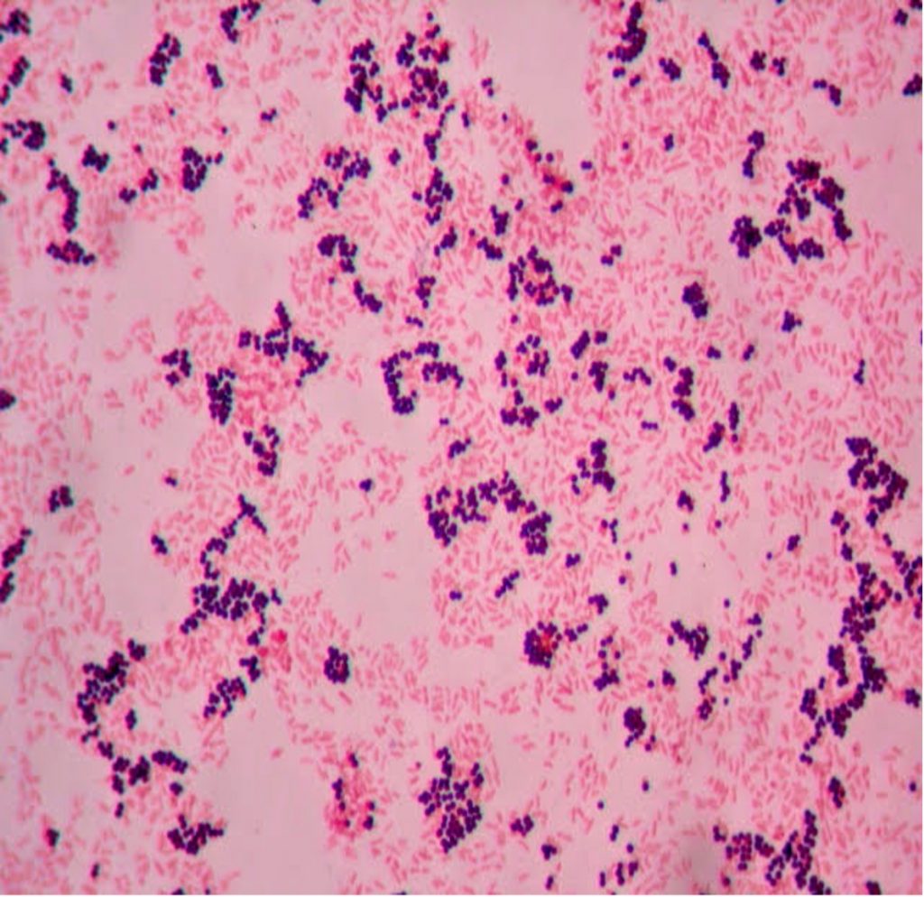
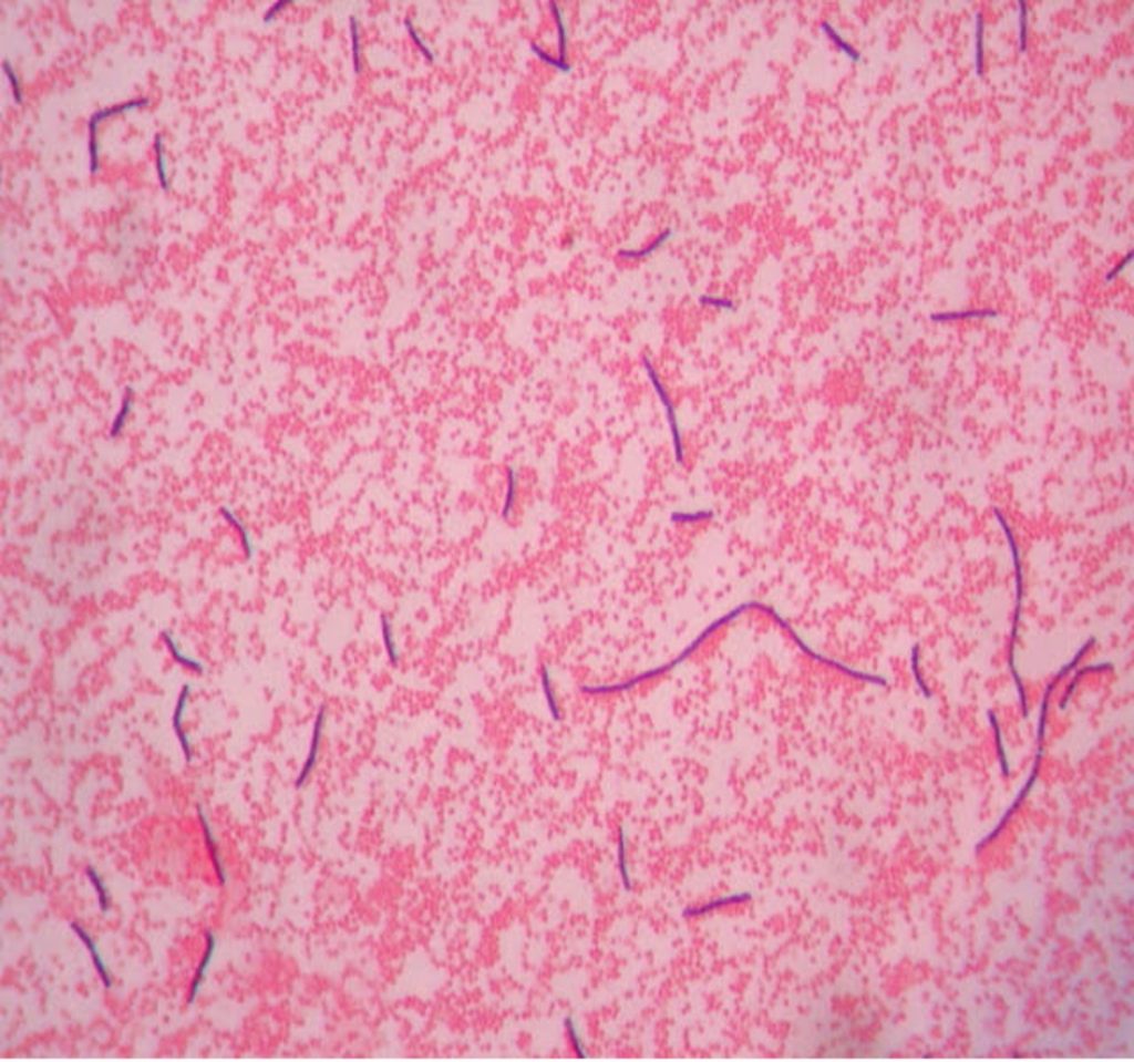
Unstained bacteria are colorless. To see them with the microscope we use chemical compounds called stains. Stains have a positive or negative charge. Because of its chemical nature, bacterial cells have a negative charge. Therefore we stain bacteria with positively charged stains. The positively charged stain is attracted to the negatively charged bacteria and the organism becomes stained. In a simple stain, the bacteria are stained with one positively charged stain, staining the bacteria the same color. In a differential stain, the bacteria are stained with two positively charged stains, staining the bacteria two colors. Simple stains provide basic information about size, morphology (shape), and arrangement. Differential stains provide information about size, shape, and arrangement, but also tells us something else about the bacteria (does the wall contain mycolic acid, are the bacteria endospore formers, the structure of the cell envelope). The Gram stain, developed by Christian Gram in 1884, is the most widely used differential stain. The Gram stain divides bacteria into two groups (Gram positive and Gram negative) based on cell envelope composition.
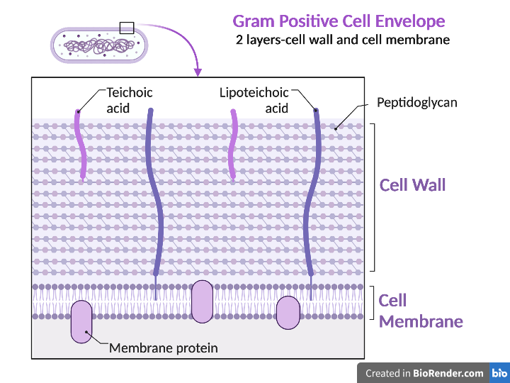
Gram positive bacteria have a cell envelope composed of two layers, a cell wall and a cell membrane. The Gram-positive cell wall is composed of hundreds of layers of peptidoglycan. The peptidoglycan layers are linked together by teichoic acids and lipoteichoic acids which anchor the peptidoglycan layers to the underlying cell membrane, respectively. The cell membrane lies underneath the thick cell wall.
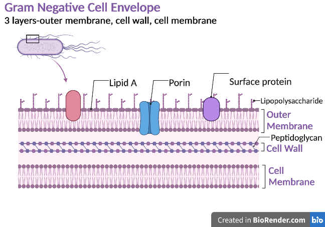
Gram negative bacteria have a cell envelope composed of three layers, an outer membrane, a cell wall, and a cell membrane. The outer membrane contains lipopolysaccharides in addition to phospholipids and proteins. The cell wall of Gram-negative bacteria is composed of just a few layers of peptidoglycan and does not contain teichoic acids. The cell membrane lies underneath the thin cell wall.
Gram stain reagents:
Crystal violet–primary stain
Gram’s Iodine–mordant that combines with crystal violet in the cell
Alcohol OR Acetone- alcohol (75% ethanol:25% acetone) –the decolorizer
Safranin–counterstain or secondary stain
The Gram stain uses four reagents. Crystal violet enters the peptidoglycan of all bacteria giving them a purple color. The next reagent is Gram’s iodine which combines with the Crystal violet to make a bigger complex in the peptidoglycan wall. The next step is the most critical. Acetone-alcohol or alcohol is used as a decolorizer which will dissolve the lipids in the outer membrane of Gram-negative cell walls. The Crystal violet-iodine complexes leak out of the thin Gram-negative cell wall. Since Gram positive cell walls lack an outer membrane, they do not decolorize and thus their thick peptidoglycan cell walls are able to retain the Crystal violet-iodine complexes. The secondary or counterstain safranin, is used to stain the Gram-negative bacteria since they lost the primary stain during decolorization. The Gram stain has proven to be very useful in the identification of bacteria and in predicting which antibiotics are most likely to be effective.
virtual Gram stain lab
Photo Requirement: Take photos or screenshots where indicated in the steps below. Paste the photos or screenshots into the Virtual Gram Stain Questions Document.
1. Go to https://virtuallabs.nmsu.edu/stain.php
2. Below this image click Continue 
3. Listen to the information on each slide in chapters 1-3. There are info and photo tabs for more information. The photos are worth the click. Click continue after you listen to each slide.
4. In Chapter 3 you will prepare a smear on a slide to Gram stain. Listen to the information and perform the tasks described on each slide in Chapter 3. Click continue after each slide.
5. In Chapter 4 you will perform the Gram stain procedure with assistance. Listen to the information and perform the tasks described on each slide in Chapter 4. Click continue after each slide.
6. In Chapter 5, you will perform the Gram stain procedure without assistance. Listen to the information and perform the tasks described on each slide in Chapter 5. You can tap the hint button if you need help remembering which step comes next. Click continue after each slide.
7. In chapter 6 the results are in. Was the yogurt contaminated with Gram positive or Gram negative bacteria? Write the answer in the Virtual Gram Stain Questions Document.
8. When you reach the end of the Virtual Gram Stain lab, take a photo or screenshot of the slide that states you have completed the Virtual Gram Stain Lab. Paste the screenshot or photo into the Virtual Gram Stain Questions Document.
