
2 Biopsychology
Any textbook on psychology would be incomplete without reference to the brain. Every behavior, thought, or experience described in the other modules must be implemented in the brain. A detailed understanding of the human brain can help us make sense of human experience and behavior. For example, one well-established fact about human cognition is that it is limited. We cannot do two complex tasks at once: We cannot read and carry on a conversation at the same time, text and drive, or surf the Internet while listening to a lecture, at least not successfully or safely. We cannot even pat our head and rub our stomach at the same time (with exceptions, see “A Brain Divided”). Why is this? Many people have suggested that such limitations reflect the fact that the behaviors draw on the same resource; if one behavior uses up most of the resource there is not enough resource left for the other. But what might this limited resource be in the brain?

The brain uses oxygen and glucose, delivered via the blood. The brain is a large consumer of these metabolites, using 20% of the oxygen and calories we consume despite being only 2% of our total weight. However, as long as we are not oxygen-deprived or malnourished, we have more than enough oxygen and glucose to fuel the brain. Thus, insufficient “brain fuel” cannot explain our limited capacity. Nor is it likely that our limitations reflect too few neurons. The average human brain contains 100 billion neurons. It is also not the case that we use only 10% of our brain, a myth that was likely started to imply we had untapped potential. Modern neuroimaging (see “Studying the Human Brain”) has shown that we use all parts of brain, just at different times, and certainly more than 10% at any one time.
If we have an abundance of brain fuel and neurons, how can we explain our limited cognitive abilities? Why can’t we do more at once? The most likely explanation is the way these neurons are wired up. We know, for instance, that many neurons in the visual cortex (the part of the brain responsible for processing visual information) are hooked up in such a way as to inhibit each other (Beck & Kastner, 2009). When one neuron fires, it suppresses the firing of other nearby neurons. If two neurons that are hooked up in an inhibitory way both fire, then neither neuron can fire as vigorously as it would otherwise. This competitive behavior among neurons limits how much visual information the brain can respond to at the same time. Similar kinds of competitive wiring among neurons may underlie many of our limitations. Thus, although talking about limited resources provides an intuitive description of our limited capacity behavior, a detailed understanding of the brain suggests that our limitations more likely reflect the complex way in which neurons talk to each other rather than the depletion of any specific resource.
Have you ever taken a device apart to find out how it works? Many of us have done so, whether to attempt a repair or simply to satisfy our curiosity. A device’s internal workings are often distinct from its user interface on the outside. For example, we don’t think about microchips and circuits when we turn up the volume on a mobile phone; instead, we think about getting the volume just right. Similarly, the inner workings of the human body are often distinct from the external expression of those workings. It is the job of psychologists to find the connection between these—for example, to figure out how the firings of millions of neurons become a thought.
This chapter strives to explain the biological mechanisms that underlie behavior. These physiological and anatomical foundations are the basis for many areas of psychology. In this chapter, you will become familiar with the structure and function of the nervous system. And, finally, you will learn how the nervous system interacts with the endocrine system.
Learning Objectives
By the end of this section, you will be able to:
- Identify the basic parts of a neuron
- Describe how neurons communicate with each other
- Explain how drugs act as agonists or antagonists for a given neurotransmitter system
Psychologists striving to understand the human mind may study the nervous system. Learning how the body’s cells and organs function can help us understand the biological basis of human psychology. The nervous system is composed of two basic cell types: glial cells (also known as glia) and neurons. Glial cells are traditionally thought to play a supportive role to neurons, both physically and metabolically. Glial cells provide scaffolding on which the nervous system is built, help neurons line up closely with each other to allow neuronal communication, provide insulation to neurons, transport nutrients, and waste products, and mediate immune responses. For years, researchers believed that there were many more glial cells than neurons; however, more recent work from Suzanna Herculano-Houzel’s laboratory has called this long-standing assumption into question and has provided important evidence that there may be a nearly 1:1 ratio of glial cells to neurons. This is important because it suggests that human brains are more similar to other primate brains than previously thought (Azevedo et al., 2009; Hercaulano-Houzel, 2012; Herculano-Houzel, 2009). Neurons, on the other hand, serve as interconnected information processors that are essential for all of the tasks of the nervous system. This section briefly describes the structure and function of neurons.
Imagine trying to string words together into a meaningful sentence without knowing the meaning of each word or its function (i.e., Is it a verb, a noun, or an adjective?). In a similar fashion, to appreciate how groups of cells work together in a meaningful way in the brain as a whole, we must first understand how individual cells in the brain function. Much like words, brain cells, called neurons, have an underlying structure that provides the foundation for their functional purpose. Have you ever seen a neuron? Did you know that the basic structure of a neuron is similar whether it is from the brain of a rat or a human? How do the billions of neurons in our brain allow us to do all the fun things we enjoy, such as texting a friend, cheering on our favorite sports team, or laughing?

Neuron Structure
Neurons are the central building blocks of the nervous system, 100 billion strong at birth. Like all cells, neurons consist of several different parts, each serving a specialized function (Figure 3.8). A neuron’s outer surface is made up of a semipermeable membrane. This membrane allows smaller molecules and molecules without an electrical charge to pass through it while stopping larger or highly charged molecules.
The nucleus of the neuron is located in the soma or cell body. The soma has branching extensions known as dendrites. The neuron is a small information processor, and dendrites serve as input sites where signals are received from other neurons. These signals are transmitted electrically across the soma and down a major extension from the soma known as the axon, which ends at multiple terminal buttons. The terminal buttons contain synaptic vesicles that house neurotransmitters, the chemical messengers of the nervous system.
Axons range in length from a fraction of an inch to several feet. In some axons, glial cells form a fatty substance known as the myelin sheath, which coats the axon and acts as an insulator, increasing the speed at which the signal travels. The myelin sheath is not continuous and there are small gaps that occur down the length of the axon. These gaps in the myelin sheath are known as the Nodes of Ranvier. The myelin sheath is crucial for the normal operation of the neurons within the nervous system: the loss of the insulation it provides can be detrimental to normal function. To understand how this works, let’s consider an example. PKU, a genetic disorder discussed earlier, causes a reduction in myelin and abnormalities in white matter cortical and subcortical structures. The disorder is associated with a variety of issues including severe cognitive deficits, exaggerated reflexes, and seizures (Anderson & Leuzzi, 2010; Huttenlocher, 2000). Another disorder, multiple sclerosis (MS), an autoimmune disorder, involves a large-scale loss of the myelin sheath on axons throughout the nervous system. The resulting interference in the electrical signal prevents the quick transmittal of information by neurons and can lead to a number of symptoms, such as dizziness, fatigue, loss of motor control, and sexual dysfunction. While some treatments may help to modify the course of the disease and manage certain symptoms, there is currently no known cure for multiple sclerosis.
In healthy individuals, the neuronal signal moves rapidly down the axon to the terminal buttons, where synaptic vesicles release neurotransmitters into the synaptic cleft (Figure 3.9). The synaptic cleft is a very small space between two neurons and is an important site where communication between neurons occurs. Once neurotransmitters are released into the synaptic cleft, they travel across it and bind with corresponding receptors on the dendrite of an adjacent neuron. Receptors, proteins on the cell surface where neurotransmitters attach, vary in shape, with different shapes “matching” different neurotransmitters.
How does a neurotransmitter “know” which receptor to bind to? The neurotransmitter and the receptor have what is referred to as a lock-and-key relationship. Specific neurotransmitters fit specific receptors similar to how a key fits a lock. The neurotransmitter binds to any receptor that it fits.
The action potential is an all-or-none phenomenon. In simple terms, this means that an incoming signal from another neuron is either sufficient or insufficient to reach the threshold of excitation. There is no in-between, and there is no turning off an action potential once it starts. Think of it as sending an email or a text message. You can think about sending it all you want, but the message is not sent until you hit the send button. Furthermore, once you send the message, there is no stopping it.
Because it is all or none, the action potential is recreated, or propagated, at its full strength at every point along the axon. Much like the lit fuse of a firecracker, it does not fade away as it travels down the axon. It is this all-or-none property that explains the fact that your brain perceives an injury to a distant body part like your toe as equally painful as one to your nose.
As noted earlier, when the action potential arrives at the terminal button, the synaptic vesicles release their neurotransmitters into the synaptic cleft. The neurotransmitters travel across the synapse and bind to receptors on the dendrites of the adjacent neuron, and the process repeats itself in the new neuron (assuming the signal is sufficiently strong to trigger an action potential). Once the signal is delivered, excess neurotransmitters in the synaptic cleft drift away, are broken down into inactive fragments or are reabsorbed in a process known as reuptake. Reuptake involves the neurotransmitter being pumped back into the neuron that released it, in order to clear the synapse (Figure 3.12). Clearing the synapse serves both to provide a clear “on” and “off” state between signals and to regulate the production of neurotransmitter (full synaptic vesicles provide signals that no additional neurotransmitters need to be produced).
Neurotransmitters and Drugs
There are several different types of neurotransmitters released by different neurons, and we can speak in broad terms about the kinds of functions associated with different neurotransmitters (Table 3.1). Much of what psychologists know about the functions of neurotransmitters come from research on the effects of drugs in psychological disorders. Psychologists who take a biological perspective and focus on the physiological causes of behavior assert that psychological disorders like depression and schizophrenia are associated with imbalances in one or more neurotransmitter systems. In this perspective, psychotropic medications can help improve the symptoms associated with these disorders. Psychotropic medications are drugs that treat psychiatric symptoms by restoring neurotransmitter balance.
| Major Neurotransmitters and How They Affect Behavior | ||
|---|---|---|
| Neurotransmitter | Involved in | Potential Effect on Behavior |
| Acetylcholine | Muscle action, memory | Increased arousal, enhanced cognition |
| Beta-endorphin | Pain, pleasure | Decreased anxiety, decreased tension |
| Dopamine | Mood, sleep, learning | Increased pleasure, suppressed appetite |
| Gamma-aminobutyric acid (GABA) | Brain function, sleep | Decreased anxiety, decreased tension |
| Glutamate | Memory, learning | Increased learning, enhanced memory |
| Norepinephrine | Heart, intestines, alertness | Increased arousal, suppressed appetite |
| Serotonin | Mood, sleep | Modulated mood, suppressed appetite |
Psychoactive drugs can act as agonists or antagonists for a given neurotransmitter system. Agonists are chemicals that mimic a neurotransmitter at the receptor site. An antagonist, on the other hand, blocks or impedes the normal activity of a neurotransmitter at the receptor. Agonists and antagonists represent drugs that are prescribed to correct the specific neurotransmitter imbalances underlying a person’s condition. For example, Parkinson’s disease, a progressive nervous system disorder, is associated with low levels of dopamine. Therefore, a common treatment strategy for Parkinson’s disease involves using dopamine agonists, which mimic the effects of dopamine by binding to dopamine receptors.
Certain symptoms of schizophrenia are associated with overactive dopamine neurotransmission. The antipsychotics used to treat these symptoms are antagonists for dopamine—they block dopamine’s effects by binding its receptors without activating them. Thus, they prevent dopamine released by one neuron from signaling information to adjacent neurons.
In contrast to agonists and antagonists, which both operate by binding to receptor sites, reuptake inhibitors prevent unused neurotransmitters from being transported back to the neuron. This allows neurotransmitters to remain active in the synaptic cleft for longer durations, increasing their effectiveness. Depression, which has been consistently linked with reduced serotonin levels, is commonly treated with selective serotonin reuptake inhibitors (SSRIs). By preventing reuptake, SSRIs strengthen the effect of serotonin, giving it more time to interact with serotonin receptors on dendrites. Common SSRIs on the market today include Prozac, Paxil, and Zoloft. The drug LSD is structurally very similar to serotonin, and it affects the same neurons and receptors as serotonin. Psychotropic drugs are not instant solutions for people suffering from psychological disorders. Often, an individual must take a drug for several weeks before seeing improvement, and many psychoactive drugs have significant negative side effects. Furthermore, individuals vary dramatically in how they respond to the drugs. To improve chances for success, it is not uncommon for people receiving pharmacotherapy to undergo psychological and/or behavioral therapies as well. Some research suggests that combining drug therapy with other forms of therapy tends to be more effective than any one treatment alone (for one such example, see March et al., 2007).
Learning Objectives
By the end of this section, you will be able to:
- Describe the difference between the central and peripheral nervous systems
- Explain the difference between the somatic and autonomic nervous systems
- Differentiate between the sympathetic and parasympathetic divisions of the autonomic nervous system
- Distinguish between gray and white matter of the cerebral hemispheres.
The mammalian nervous system is a complex biological organ, which enables many animals including humans to function in a coordinated fashion. The original design of this system is preserved across many animals through evolution; thus, adaptive physiological and behavioral functions are similar across many animal species. Comparative study of physiological functioning in the nervous systems of different animals lend insights to their behavior and their mental processing and make it easier for us to understand the human brain and behavior. In addition, studying the development of the nervous system in a growing human provides a wealth of information about the change in its form and behaviors that result from this change. The nervous system is divided into central and peripheral nervous systems, and the two heavily interact with one another. The peripheral nervous system controls volitional (somatic nervous system) and nonvolitional (autonomic nervous system) behaviors using cranial and spinal nerves. The central nervous system is divided into forebrain, midbrain, and hindbrain, and each division performs a variety of tasks; for example, the cerebral cortex in the forebrain houses sensory, motor, and associative areas that gather sensory information, process information for perception and memory, and produce responses based on incoming and inherent information. To study the nervous system, a number of methods have evolved over time; these methods include examining brain lesions, microscopy, electrophysiology, electroencephalography, and many scanning technologies.
Evolution of the Nervous System
Many scientists and thinkers (Cajal, 1937; Crick & Koch, 1990; Edelman, 2004) believe that the human nervous system is the most complex machine known to man. Its complexity points to one undeniable fact—that it has evolved slowly over time from simpler forms. Evolution of the nervous system is intriguing not because we can marvel at this complicated biological structure, but it is fascinating because it inherits a lineage of a long history of many less complex nervous systems (Figure 1), and it documents a record of adaptive behaviors observed in life forms other than humans. Thus, evolutionary study of the nervous system is important, and it is the first step in understanding its design, its workings, and its functional interface with the environment.

The brains of some animals, like apes, monkeys, and rodents, are structurally similar to humans (Figure 1), while others are not (e.g., invertebrates, single-celled organisms). Does anatomical similarity of these brains suggest that behaviors that emerge in these species are also similar? Indeed, many animals display behaviors that are similar to humans, e.g., apes use nonverbal communication signals with their hands and arms that resemble nonverbal forms of communication in humans (Gardner & Gardner, 1969; Goodall, 1986; Knapp & Hall, 2009). If we study very simple behaviors, like physiological responses made by individual neurons, then brain-based behaviors of invertebrates (Kandel & Schwartz, 1982) look very similar to humans, suggesting that from time immemorial such basic behaviors have been conserved in the brains of many simple animal forms and in fact are the foundation of more complex behaviors in animals that evolved later (Bullock, 1984).
Even at the micro-anatomical level, we note that individual neurons differ in complexity across animal species. Human neurons exhibit more intricate complexity than other animals; for example, neuronal processes (dendrites) in humans have many more branch points, branches, and spines.
Complexity in the structure of the nervous system, both at the macro- and micro-levels, give rise to complex behaviors. We can observe similar movements of the limbs, as in nonverbal communication, in apes and humans, but the variety and intricacy of nonverbal behaviors using hands in humans surpasses apes. Deaf individuals who use American Sign Language (ASL) express themselves in English nonverbally; they use this language with such fine gradation that many accents of ASL exist (Walker, 1987). Complexity of behavior with increasing complexity of the nervous system, especially the cerebral cortex, can be observed in the genus Homo (Figure 2). If we compare sophistication of material culture in Homo habilis (2 million years ago; brain volume ~650 cm3) and Homo sapiens (300,000 years to now; brain volume ~1400 cm3), the evidence shows that Homo habilis used crude stone tools compared with modern tools used by Homo sapiens to erect cities, develop written languages, embark on space travel, and study her own self. All of this is due to increasing complexity of the nervous system.

What has led to the complexity of the brain and nervous system through evolution, to its behavioral and cognitive refinement? Darwin (1859, 1871) proposed two forces of natural and sexual selection as work engines behind this change. He prophesied, “psychology will be based on a new foundation, that of the necessary acquirement of each mental power and capacity by gradation” that is, psychology will be based on evolution (Rosenzweig, Breedlove, & Leiman, 2002).
Development of the Nervous System
Where the study of change in the nervous system over eons is immensely captivating, studying the change in a single brain during individual development is no less engaging. In many ways the ontogeny (development) of the nervous system in an individual mimics the evolutionary advancement of this structure observed across many animal species. During development, the nervous tissue emerges from the ectoderm (one of the three layers of the mammalian embryo) through the process of neural induction. This process causes the formation of the neural tube, which extends in a rostrocaudal (head-to-tail) plane. The tube, which is hollow, seams itself in the rostrocaudal direction. In some disease conditions, the neural tube does not close caudally and results in an abnormality called spina bifida. In this pathological condition, the lumbar and sacral segments of the spinal cord are disrupted.
As gestation progresses, the neural tube balloons up (cephalization) at the rostral end, and forebrain, midbrain, hindbrain, and the spinal cord can be visually delineated (day 40). About 50 days into gestation, six cephalic areas can be anatomically discerned (also see below for a more detailed description of these areas).
The progenitor cells (neuroblasts) that form the lining (neuroepithelium) of the neural tube generate all the neurons and glial cells of the central nervous system. During early stages of this development, neuroblasts rapidly divide and specialize into many varieties of neurons and glial cells, but this proliferation of cells is not uniform along the neural tube—that is why we see the forebrain and hindbrain expand into larger cephalic tissues than the midbrain. The neuroepithelium also generates a group of specialized cells that migrate outside the neural tube to form the neural crest. This structure gives rise to sensory and autonomic neurons in the peripheral nervous system.
The Structure of the Nervous System
The mammalian nervous system is divided into central and peripheral nervous systems.
The Peripheral Nervous System

The peripheral nervous system is divided into somatic and autonomic nervous systems (Figure 3). Where the somatic nervous system consists of cranial nerves (12 pairs) and spinal nerves (31 pairs) and is under the volitional control of the individual in maneuvering bodily muscles, the autonomic nervous system also running through these nerves lets the individual have little control over muscles and glands. Main divisions of the autonomic nervous system that control visceral structures are the sympathetic and parasympathetic nervous systems.
At an appropriate cue (say a fear-inducing object like a snake), the sympathetic division generally energizes many muscles (e.g., heart) and glands (e.g., adrenals), causing activity and release of hormones that lead the individual to negotiate the fear-causing snake with fight-or-flight responses. Whether the individual decides to fight the snake or run away from it, either action requires energy; in short, the sympathetic nervous system says “go, go, go.” The parasympathetic nervous system, on the other hand, curtails undue energy mobilization into muscles and glands and modulates the response by saying “stop, stop, stop.” This push–pull tandem system regulates fight-or-flight responses in all of us.
While it is clear that such a response would be critical for survival for our ancestors, who lived in a world full of real physical threats, many of the high-arousal situations we face in the modern world are more psychological in nature. For example, think about how you feel when you have to stand up and give a presentation in front of a roomful of people, or right before taking a big test. You are in no real physical danger in those situations, and yet you have evolved to respond to a perceived threat with the fight or flight response. This kind of response is not nearly as adaptive in the modern world; in fact, we suffer negative health consequences when faced constantly with psychological threats that we can neither fight nor flee. Recent research suggests that an increase in susceptibility to heart disease (Chandola, Brunner, & Marmot, 2006) and impaired function of the immune system (Glaser & Kiecolt-Glaser, 2005) are among the many negative consequences of persistent and repeated exposure to stressful situations. Some of this tendency for stress reactivity can be wired by early experiences of trauma.
Once the threat has been resolved, the parasympathetic nervous system takes over and returns bodily functions to a relaxed state. Our hunter’s heart rate and blood pressure return to normal, his pupils constrict, he regains control of his bladder, and the liver begins to store glucose in the form of glycogen for future use. These restorative processes are associated with the activation of the parasympathetic nervous system.
The Central Nervous System
, Cerebrum (Telencephalon), and the Cerebrum, is in turn comprised of Isocortex, the Limbic System and Basal Ganglia. The Midbrain or Mesencephalon is composed of Superior and Inferior Colliculi and the Reticular Activation System. The Hindbrain, or Rhombencephalon contains Medulla, Pons and Cerebellum with the latter two being a part of the Metencephalon.")
The central nervous system is divided into a number of important parts (see Figure 4), including the spinal cord, each specialized to perform a set of specific functions. Telencephalon or cerebrum is a newer development in the evolution of the mammalian nervous system. In humans, it is about the size of a large napkin and when crumpled into the skull, it forms furrows called sulci (singular form, sulcus). The bulges between sulci are called gyri (singular form, gyrus). The cortex is divided into two hemispheres, and each hemisphere is further divided into four lobes (Figure 5a), which have specific functions. The division of these lobes is based on two delineating sulci: the central sulcus divides the hemisphere into frontal and parietal-occipital lobes and the lateral sulcus marks the temporal lobe, which lies below.

Just in front of the central sulcus lies an area called the primary motor cortex (precentral gyrus), which connects to the muscles of the body, and on volitional command moves them. From mastication to movements in the genitalia, the body map is represented on this strip (Figure 5b).
Some body parts, like fingers, thumbs, and lips, occupy a greater representation on the strip than, say, the trunk. This disproportionate representation of the body on the primary motor cortex is called the magnification factor (Rolls & Cowey, 1970) and is seen in other motor and sensory areas. At the lower end of the central sulcus, close to the lateral sulcus, lies the Broca’s area (Figure 6b) in the left frontal lobe, which is involved with language production. Damage to this part of the brain led Pierre Paul Broca, a French neuroscientist in 1861, to document many different forms of aphasias, in which his patients would lose the ability to speak or would retain partial speech impoverished in syntax and grammar (AAAS, 1880). It is no wonder that others have found subvocal rehearsal and central executive processes of working memory in this frontal lobe (Smith & Jonides, 1997, 1999).

Just behind the central gyrus, in the parietal lobe, lies the primary somatosensory cortex (Figure 6a) on the postcentral gyrus, which represents the whole body receiving inputs from the skin and muscles. The primary somatosensory cortex parallels, abuts, and connects heavily to the primary motor cortex and resembles it in terms of areas devoted to bodily representation. All spinal and some cranial nerves (e.g., the facial nerve) send sensory signals from skin (e.g., touch) and muscles to the primary somatosensory cortex. Close to the lower (ventral) end of this strip, curved inside the parietal lobe, is the taste area (secondary somatosensory cortex), which is involved with taste experiences that originate from the tongue, pharynx, epiglottis, and so forth.

Just below the parietal lobe, and under the caudal end of the lateral fissure, in the temporal lobe, lies the Wernicke’s area (Demonet et al., 1992). This area is involved with language comprehension and is connected to the Broca’s area through the arcuate fasciculus, nerve fibers that connect these two regions. Damage to the Wernicke’s area (Figure 6b) results in many kinds of agnosias; agnosia is defined as an inability to know or understand language and speech-related behaviors. So an individual may show word deafness, which is an inability to recognize spoken language, or word blindness, which is an inability to recognize written or printed language. Close in proximity to the Wernicke’s area is the primary auditory cortex, which is involved with audition, and finally the brain region devoted to smell (olfaction) is tucked away inside the primary olfactory cortex (prepyriform cortex).

At the very back of the cerebral cortex lies the occipital lobe housing the primary visual cortex. Optic nerves travel all the way to the thalamus (lateral geniculate nucleus, LGN) and then to visual cortex, where images that are received on the retina are projected (Hubel, 1995).
In the past 50 to 60 years, visual sense and visual pathways have been studied extensively, and our understanding about them has increased manifold. We now understand that all objects that form images on the retina are transformed (transduction) in neural language handed down to the visual cortex for further processing. In the visual cortex, all attributes (features) of the image, such as the color, texture, and orientation, are decomposed and processed by different visual cortical modules (Van Essen, Anderson & Felleman, 1992) and then recombined to give rise to singular perception of the image in question.
If we cut the cerebral hemispheres in the middle, a new set of structures come into view. Many of these perform different functions vital to our being. For example, the limbic system contains a number of nuclei that process memory (hippocampus and fornix) and attention and emotions (cingulate gyrus); the globus pallidus is involved with motor movements and their coordination; the hypothalamus and thalamus are involved with drives, motivations, and trafficking of sensory and motor throughputs. The hypothalamus plays a key role in regulating endocrine hormones in conjunction with the pituitary gland that extends from the hypothalamus through a stalk (infundibulum).

As we descend down the thalamus, the midbrain comes into view with superior and inferior colliculi, which process visual and auditory information, as does the substantia nigra, which is involved with notorious Parkinson’s disease, and the reticular formation regulating arousal, sleep, and temperature. A little lower, the hindbrain with the pons processes sensory and motor information employing the cranial nerves, works as a bridge that connects the cerebral cortex with the medulla, and reciprocally transfers information back and forth between the brain and the spinal cord. The medulla oblongata processes breathing, digestion, heart and blood vessel function, swallowing, and sneezing. The cerebellum controls motor movement coordination, balance, equilibrium, and muscle tone.
The midbrain and the hindbrain, which make up the brain stem, culminate in the spinal cord. Whereas inside the cerebral cortex, the gray matter (neuronal cell bodies) lies outside and white matter (myelinated axons) inside; in the spinal cord this arrangement reverses, as the gray matter resides inside and the white matter outside. Paired nerves (ganglia) exit the spinal cord, some closer in direction towards the back (dorsal) and others towards the front (ventral). The dorsal nerves (afferent) receive sensory information from skin and muscles, and ventral nerves (efferent) send signals to muscles and organs to respond.
Gray Versus White Matter
The cerebral hemispheres contain both grey and white matter, so called because they appear grayish and whitish in dissections or in an MRI (magnetic resonance imaging; see, “Studying the Human Brain”). The gray matter is composed of the neuronal cell bodies (see module, “Neurons”). The cell bodies (or soma) contain the genes of the cell and are responsible for metabolism (keeping the cell alive) and synthesizing proteins. In this way, the cell body is the workhorse of the cell. The white matter is composed of the axons of the neurons, and, in particular, axons that are covered with a sheath of myelin (fatty support cells that are whitish in color). Axons conduct the electrical signals from the cell and are, therefore, critical to cell communication. People use the expression “use your gray matter” when they want a person to think harder. The “gray matter” in this expression is probably a reference to the cerebral hemispheres more generally; the gray cortical sheet (the convoluted surface of the cortex) being the most visible. However, both the gray matter and white matter are critical to proper functioning of the mind. Losses of either result in deficits in language, memory, reasoning, and other mental functions. See Figure 3 for MRI slices showing both the inner white matter that connects the cell bodies in the gray cortical sheet.
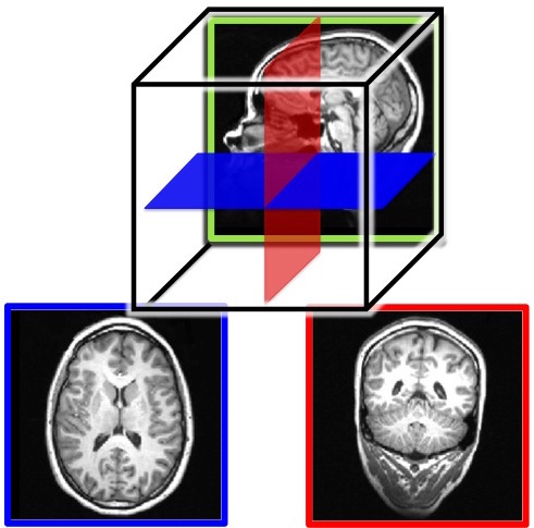
Figure 3. MRI slices of the human brain. Both the outer gray matter and inner white matter are visible in each image. The brain is a three-dimensional (3-D) structure, but an image is two-dimensional (2-D). Here, we show example slices of the three possible 2-D cuts through the brain: a saggital slice (top image), a horizontal slice (bottom left), which is also known as a transverse or axial slice, and a coronal slice (bottom right). The bottom two images are color-coded to match the illustration of the relative orientations of the three slices in the top image.
Studying the Nervous System
The study of the nervous system involves anatomical and physiological techniques that have improved over the years in efficiency and caliber. Clearly, gross morphology of the nervous system requires an eye-level view of the brain and the spinal cord. However, to resolve minute components, optical and electron microscopic techniques are needed.
Light microscopes and, later, electron microscopes have changed our understanding of the intricate connections that exist among nerve cells. For example, modern staining procedures (immunocytochemistry) make it possible to see selected neurons that are of one type or another or are affected by growth. With better resolution of the electron microscopes, fine structures like the synaptic cleft between the pre- and post-synaptic neurons can be studied in detail. Along with the neuroanatomical techniques, a number of other methodologies aid neuroscientists in studying the function and physiology of the nervous system. These methods will be explored later on in the chapter.
Understanding the nervous system has been a long journey of inquiry, spanning several hundreds of years of meticulous studies carried out by some of the most creative and versatile investigators in the fields of philosophy, evolution, biology, physiology, anatomy, neurology, neuroscience, cognitive sciences, and psychology. Despite our profound understanding of this organ, its mysteries continue to surprise us, and its intricacies make us marvel at this complex structure unmatched in the universe.
Learning Objectives
By the end of this section, you will be able to:
- Explain the functions of the spinal cord
- Identify the hemispheres of the brain
- Name and describe the basic function of the four cerebral lobes: occipital, temporal, parietal, and frontal cortex.
- Describe a split-brain patient and at least two important aspects of brain function that these patients reveal.
The brain is a remarkably complex organ comprised of billions of interconnected neurons and glia. It is a bilateral, or two-sided, structure that can be separated into distinct lobes. Each lobe is associated with certain types of functions, but, ultimately, all of the areas of the brain interact with one another to provide the foundation for our thoughts and behaviors. In this section, we discuss the overall organization of the brain and the functions associated with different brain areas, beginning with what can be seen as an extension of the brain, the spinal cord.
The Spinal Cord
It can be said that the spinal cord is what connects the brain to the outside world. Because of it, the brain can act. The spinal cord is like a relay station, but a very smart one. It not only routes messages to and from the brain, but it also has its own system of automatic processes, called reflexes.
The top of the spinal cord is a bundle of nerves that merges with the brain stem, where the basic processes of life are controlled, such as breathing and digestion. In the opposite direction, the spinal cord ends just below the ribs—contrary to what we might expect, it does not extend all the way to the base of the spine.
The spinal cord is functionally organized in 30 segments, corresponding with the vertebrae. Each segment is connected to a specific part of the body through the peripheral nervous system. Nerves branch out from the spine at each vertebra. Sensory nerves bring messages in; motor nerves send messages out to the muscles and organs. Messages travel to and from the brain through every segment.
Some sensory messages are immediately acted on by the spinal cord, without any input from the brain. Withdrawal from a hot object and the knee jerk are two examples. When a sensory message meets certain parameters, the spinal cord initiates an automatic reflex. The signal passes from the sensory nerve to a simple processing center, which initiates a motor command. Seconds are saved because messages don’t have to go the brain, be processed, and get sent back. In matters of survival, the spinal reflexes allow the body to react extraordinarily fast.
The spinal cord is protected by bony vertebrae and cushioned in cerebrospinal fluid, but injuries still occur. When the spinal cord is damaged in a particular segment, all lower segments are cut off from the brain, causing paralysis. Therefore, the lower on the spine damage occurs, the fewer functions an injured individual will lose.
Neuroplasticity
Bob Woodruff, a reporter for ABC, suffered a traumatic brain injury after a bomb exploded next to the vehicle he was in while covering a news story in Iraq. As a consequence of these injuries, Woodruff experienced many cognitive deficits including difficulties with memory and language. However, over time and with the aid of intensive amounts of cognitive and speech therapy, Woodruff has shown an incredible recovery of function (Fernandez, 2008, October 16).
One of the factors that made this recovery possible was neuroplasticity. Neuroplasticity refers to how the nervous system can change and adapt. Neuroplasticity can occur in a variety of ways including personal experiences, developmental processes, or, as in Woodruff’s case, in response to some sort of damage or injury that has occurred. Neuroplasticity can involve the creation of new synapses, pruning of synapses that are no longer used, changes in glial cells, and even the birth of new neurons. Because of neuroplasticity, our brains are constantly changing and adapting, and while our nervous system is most plastic when we are very young, as Woodruff’s case suggests, it is still capable of remarkable changes later in life.
The Two Hemispheres
The surface of the brain, known as the cerebral cortex, is very uneven, characterized by a distinctive pattern of folds or bumps, known as gyri (singular: gyrus), and grooves, known as sulci (singular: sulcus), shown in Figure 3.15. These gyri and sulci form important landmarks that allow us to separate the brain into functional centers. The most prominent sulcus, known as the longitudinal fissure, is the deep groove that separates the brain into two halves or hemispheres: the left hemisphere and the right hemisphere.
There is evidence of specialization of function—referred to as lateralization—in each hemisphere, mainly regarding differences in language functions. The left hemisphere controls the right half of the body, and the right hemisphere controls the left half of the body. Decades of research on lateralization of function by Michael Gazzaniga and his colleagues suggest that a variety of functions ranging from cause-and-effect reasoning to self-recognition may follow patterns that suggest some degree of hemispheric dominance (Gazzaniga, 2005). For example, the left hemisphere has been shown to be superior for forming associations in memory, selective attention, and positive emotions. The right hemisphere, on the other hand, has been shown to be superior in pitch perception, arousal, and negative emotions (Ehret, 2006). However, it should be pointed out that research on which hemisphere is dominant in a variety of different behaviors has produced inconsistent results, and therefore, it is probably better to think of how the two hemispheres interact to produce a given behavior rather than attributing certain behaviors to one hemisphere versus the other (Banich & Heller, 1998).
The two hemispheres are connected by a thick band of neural fibers known as the corpus callosum, consisting of about 200 million axons. The corpus callosum allows the two hemispheres to communicate with each other and allows for information being processed on one side of the brain to be shared with the other side.
Normally, we are not aware of the different roles that our two hemispheres play in day-to-day functions, but there are people who come to know the capabilities and functions of their two hemispheres quite well. In some cases of severe epilepsy, doctors elect to sever the corpus callosum as a means of controlling the spread of seizures (Figure 3.16). While this is an effective treatment option, it results in individuals who have “split brains.” After surgery, these split-brain patients show a variety of interesting behaviors. For instance, a split-brain patient is unable to name a picture that is shown in the patient’s left visual field because the information is only available in the largely nonverbal right hemisphere. However, they are able to recreate the picture with their left hand, which is also controlled by the right hemisphere. When the more verbal left hemisphere sees the picture that the hand drew, the patient is able to name it (assuming the left hemisphere can interpret what was drawn by the left hand).
Much of what we know about the functions of different areas of the brain comes from studying changes in the behavior and ability of individuals who have suffered damage to the brain. For example, researchers study the behavioral changes caused by strokes to learn about the functions of specific brain areas. A stroke, caused by an interruption of blood flow to a region in the brain, causes a loss of brain function in the affected region. The damage can be in a small area, and, if it is, this gives researchers the opportunity to link any resulting behavioral changes to a specific area. The types of deficits displayed after a stroke will be largely dependent on where in the brain the damage occurred.
Consider Theona, an intelligent, self-sufficient woman, who is 62 years old. Recently, she suffered a stroke in the front portion of her right hemisphere. As a result, she has great difficulty moving her left leg. (As you learned earlier, the right hemisphere controls the left side of the body; also, the brain’s main motor centers are located at the front of the head, in the frontal lobe.) Theona has also experienced behavioral changes. For example, while in the produce section of the grocery store, she sometimes eats grapes, strawberries, and apples directly from their bins before paying for them. This behavior—which would have been very embarrassing to her before the stroke—is consistent with damage in another region in the frontal lobe—the prefrontal cortex, which is associated with judgment, reasoning, and impulse control.
Forebrain Structures
The two hemispheres of the cerebral cortex are part of the forebrain (Figure 3.17), which is the largest part of the brain. The forebrain contains the cerebral cortex and a number of other structures that lie beneath the cortex (called subcortical structures): thalamus, hypothalamus, pituitary gland, and the limbic system (a collection of structures). The cerebral cortex, which is the outer surface of the brain, is associated with higher-level processes such as consciousness, thought, emotion, reasoning, language, and memory. Each cerebral hemisphere can be subdivided into four lobes, each associated with different functions.
Lobes of the Brain
The four lobes of the brain are the frontal, parietal, temporal, and occipital lobes (Figure 3.18). The frontal lobe is located in the forward part of the brain, extending back to a fissure known as the central sulcus. The frontal lobe is involved in reasoning, motor control, emotion, and language. It contains the motor cortex, which is involved in planning and coordinating movement; the prefrontal cortex, which is responsible for higher-level cognitive functioning; and Broca’s area, which is essential for language production.
People who suffer damage to Broca’s area have great difficulty producing language of any form (Figure 3.18). For example, Padma was an electrical engineer who was socially active and a caring, involved parent. About twenty years ago, she was in a car accident and suffered damage to her Broca’s area. She completely lost the ability to speak and form any kind of meaningful language. There is nothing wrong with her mouth or her vocal cords, but she is unable to produce words. She can follow directions but can’t respond verbally, and she can read but no longer write. She can do routine tasks like running to the market to buy milk, but she could not communicate verbally if a situation called for it.
Probably the most famous case of frontal lobe damage is that of a man by the name of Phineas Gage. On September 13, 1848, Gage (age 25) was working as a railroad foreman in Vermont. He and his crew were using an iron rod to tamp explosives down into a blasting hole to remove rock along the railway’s path. Unfortunately, the iron rod created a spark and caused the rod to explode out of the blasting hole, into Gage’s face, and through his skull (Figure 3.19). Although lying in a pool of his own blood with brain matter emerging from his head, Gage was conscious and able to get up, walk, and speak.
However, there is some debate on what long-term effects Gage experienced after the accident. Gage’s case occurred in the midst of a 19th-century debate over localization—regarding whether certain areas of the brain are associated with particular functions. On the basis of extremely limited information about Gage, the extent of his injury, and his life before and after the accident, scientists tended to find support for their own views, on whichever side of the debate they fell (Macmillan, 1999). What we can conclude from his accident is that he was able to live a full life after his brain injury and that the brain is incredibly resilient.
The brain’s parietal lobe is located immediately behind the frontal lobe and is involved in processing information from the body’s senses. It contains the somatosensory cortex, which is essential for processing sensory information from across the body, such as touch, temperature, and pain. The somatosensory cortex is organized topographically, which means that spatial relationships that exist in the body are generally maintained on the surface of the somatosensory cortex (Figure 3.20). For example, the portion of the cortex that processes sensory information from the hand is adjacent to the portion that processes information from the wrist.
The temporal lobe is located on the side of the head (temporal means “near the temples”), and is associated with hearing, memory, emotion, and some aspects of language. The auditory cortex, the main area responsible for processing auditory information, is located within the temporal lobe. Wernicke’s area, important for speech comprehension, is also located here. Whereas individuals with damage to Broca’s area have difficulty producing language, those with damage to Wernicke’s area can produce sensible language, but they are unable to understand it (Figure 3.21).
The occipital lobe is located at the very back of the brain and contains the primary visual cortex, which is responsible for interpreting incoming visual information. The occipital cortex is organized retinotopically, which means there is a close relationship between the position of an object in a person’s visual field and the position of that object’s representation on the cortex. You will learn much more about how visual information is processed in the occipital lobe when you study sensation and perception.
Other Areas of the Forebrain
Other areas of the forebrain, located beneath the cerebral cortex, include the thalamus and the limbic system. The thalamus is a sensory relay for the brain. All of our senses, with the exception of smell, are routed through the thalamus before being directed to other areas of the brain for processing (Figure 3.22).
The limbic system is involved in processing both emotion and memory. Interestingly, the sense of smell projects directly to the limbic system; therefore, not surprisingly, the sense of smell can evoke emotional responses in ways that other sensory modalities cannot. The limbic system is made up of a number of different structures, but three of the most important are the hippocampus, the amygdala, and the hypothalamus (Figure 3.23). The hippocampus is an essential structure for learning and memory. The amygdala is involved in our experience of emotion and in tying emotional meaning to our memories. The hypothalamus regulates a number of homeostatic processes, including the regulation of body temperature, appetite, and blood pressure. The hypothalamus also serves as an interface between the nervous system and the endocrine system and in the regulation of sexual motivation and behavior.
The Case of Henry Molaison (H.M.)
In 1953, Henry Gustav Molaison (H. M.) was a 27-year-old man who experienced severe seizures. In an attempt to control his seizures, H. M. underwent brain surgery to remove his hippocampus and amygdala. Following the surgery, H.M’s seizures became much less severe, but he also suffered some unexpected—and devastating—consequences of the surgery: he lost his ability to form many types of new memories. For example, he was unable to learn new facts, such as who was president of the United States. He was able to learn new skills, but afterward, he had no recollection of learning them. For example, while he might learn to use a computer, he would have no conscious memory of ever having used one. He could not remember new faces, and he was unable to remember events, even immediately after they occurred. Researchers were fascinated by his experience, and he is considered one of the most studied cases in medical and psychological history (Hardt, Einarsson, & Nader, 2010; Squire, 2009). Indeed, his case has provided tremendous insight into the role that the hippocampus plays in the consolidation of new learning into explicit memory.
Midbrain and Hindbrain Structures
The midbrain is comprised of structures located deep within the brain, between the forebrain and the hindbrain. The reticular formation is centered in the midbrain, but it actually extends up into the forebrain and down into the hindbrain. The reticular formation is important in regulating the sleep/wake cycle, arousal, alertness, and motor activity.
The substantia nigra (Latin for “black substance”) and the ventral tegmental area (VTA) are also located in the midbrain (Figure 3.24). Both regions contain cell bodies that produce the neurotransmitter dopamine, and both are critical for movement. Degeneration of the substantia nigra and VTA is involved in Parkinson’s disease. In addition, these structures are involved in mood, reward, and addiction (Berridge & Robinson, 1998; Gardner, 2011; George, Le Moal, & Koob, 2012).
The hindbrain is located at the back of the head and looks like an extension of the spinal cord. It contains the medulla, pons, and cerebellum (Figure 3.25). The medulla controls the automatic processes of the autonomic nervous system, such as breathing, blood pressure, and heart rate. The word pons literally means “bridge,” and as the name suggests, the pons serves to connect the hindbrain to the rest of the brain. It also is involved in regulating brain activity during sleep. The medulla, pons, and various structures are known as the brainstem, and aspects of the brainstem span both the midbrain and the hindbrain.
The cerebellum (Latin for “little brain”) receives messages from muscles, tendons, joints, and structures in our ear to control balance, coordination, movement, and motor skills. The cerebellum is also thought to be an important area for processing some types of memories. In particular, procedural memory, or memory involved in learning and remembering how to perform tasks, is thought to be associated with the cerebellum. Recall that H. M. was unable to form new explicit memories, but he could learn new tasks. This is likely due to the fact that H. M.’s cerebellum remained intact.
Learning Objectives
- Name and describe the most common approaches to studying the human brain.
- Distinguish among neuroimaging methods
Studying the Human Brain
How do we know what the brain does? We have gathered knowledge about the functions of the brain from many different methods. Each method is useful for answering distinct types of questions, but the strongest evidence for a specific role or function of a particular brain area is converging evidence; that is, similar findings reported from multiple studies using different methods.
One of the first organized attempts to study the functions of the brain was phrenology, a popular field of study in the first half of the 19th century. Phrenologists assumed that various features of the brain, such as its uneven surface, are reflected on the skull; therefore, they attempted to correlate bumps and indentations of the skull with specific functions of the brain. For example, they would claim that a very artistic person has ridges on the head that vary in size and location from those of someone who is very good at spatial reasoning. Although the assumption that the skull reflects the underlying brain structure has been proven wrong, phrenology nonetheless significantly impacted current-day neuroscience and its thinking about the functions of the brain. That is, different parts of the brain are devoted to very specific functions that can be identified through scientific inquiry.
Neuroanatomy
Dissection of the brain, in either animals or cadavers, has been a critical tool of neuroscientists since 340 BC when Aristotle first published his dissections. Since then this method has advanced considerably with the discovery of various staining techniques that can highlight particular cells. Because the brain can be sliced very thinly, examined under the microscope, and particular cells highlighted, this method is especially useful for studying specific groups of neurons or small brain structures; that is, it has a very high spatial resolution. Dissections allow scientists to study changes in the brain that occur due to various diseases or experiences (e.g., exposure to drugs or brain injuries).
Virtual dissection studies with living humans are also conducted. Here, the brain is imaged using computerized axial tomography (CAT) or MRI scanners; they reveal with very high precision the various structures in the brain and can help detect changes in gray or white matter. These changes in the brain can then be correlated with behavior, such as performance on memory tests, and, therefore, implicate specific brain areas in certain cognitive functions.
Some researchers induce lesions or ablate (i.e., remove) parts of the brain in animals. If the animal’s behavior changes after the lesion, we can infer that the removed structure is important for that behavior. Lesions of human brains are studied in patient populations only; that is, patients who have lost a brain region due to a stroke or other injury, or who have had surgical removal of a structure to treat a particular disease (e.g., a callosotomy to control epilepsy, as in split-brain patients). From such case studies, we can infer brain function by measuring changes in the behavior of the patients before and after the lesion.
Neuroimaging
You have learned how brain injury can provide information about the functions of different parts of the brain. Increasingly, however, we are able to obtain that information using brain imaging techniques on individuals who have not suffered a brain injury. In this section, we take a more in-depth look at some of the techniques that are available for imaging the brain, including techniques that rely on radiation, magnetic fields, or electrical activity within the brain.
Techniques Involving Radiation
A computerized tomography (CT) scan involves taking a number of x-rays of a particular section of a person’s body or brain (Figure 3.26). The x-rays pass through tissues of different densities at different rates, allowing a computer to construct an overall image of the area of the body being scanned. A CT scan is often used to determine whether someone has a tumor or significant brain atrophy.
Positron emission tomography (PET) scans create pictures of the living, active brain (Figure 3.27). An individual receiving a PET scan drinks or is injected with a mildly radioactive substance called a tracer. Once in the bloodstream, the amount of tracer in any given region of the brain can be monitored. As a brain area becomes more active, more blood flows to that area. A computer monitors the movement of the tracer and creates a rough map of active and inactive areas of the brain during a given behavior. PET scans show little detail, are unable to pinpoint events precisely in time, and require that the brain be exposed to radiation; therefore, this technique has been replaced by the fMRI as an alternative diagnostic tool. However, combined with CT, PET technology is still being used in certain contexts. For example, CT/PET scans allow better imaging of the activity of neurotransmitter receptors and open new avenues in schizophrenia research. In this hybrid CT/PET technology, CT contributes clear images of brain structures, while PET shows the brain’s activity.
Techniques Involving Magnetic Fields
In magnetic resonance imaging (MRI), a person is placed inside a machine that generates a strong magnetic field. The magnetic field causes the hydrogen atoms in the body’s cells to move. When the magnetic field is turned off, the hydrogen atoms emit electromagnetic signals as they return to their original positions. Tissues of different densities give off different signals, which a computer interprets and displays on a monitor. Functional magnetic resonance imaging (fMRI) operates on the same principles, but it shows changes in brain activity over time by tracking blood flow and oxygen levels. The fMRI provides more detailed images of the brain’s structure, as well as better accuracy in time than is possible in PET scans (Figure 3.28). With their high level of detail, MRI and fMRI are often used to compare the brains of healthy individuals to the brains of individuals diagnosed with psychological disorders. This comparison helps determine what structural and functional differences exist between these populations.

Techniques Involving Electrical Activity
In some situations, it is helpful to gain an understanding of the overall activity of a person’s brain, without needing information on the actual location of the activity. Electroencephalography (EEG) serves this purpose by providing a measure of a brain’s electrical activity. An array of electrodes is placed around a person’s head (Figure 3.29). The signals received by the electrodes result in a printout of the electrical activity of his or her brain, or brainwaves, showing both the frequency (number of waves per second) and amplitude (height) of the recorded brainwaves, with an accuracy within milliseconds. Such information is especially helpful to researchers studying sleep patterns among individuals with sleep disorders.
Learning Objectives
By the end of this section, you will be able to:
- Identify the major glands of the endocrine system
- Identify the hormones secreted by each gland
- Describe each hormone’s role in regulating bodily functions
This module describes the relationship between hormones and behavior. Many readers are likely already familiar with the general idea that hormones can affect behavior. Students are generally familiar with the idea that sex-hormone concentrations increase in the blood during puberty and decrease as we age, especially after about 50 years of age. Sexual behavior shows a similar pattern. Most people also know about the relationship between aggression and anabolic steroid hormones, and they know that administration of artificial steroid hormones sometimes results in uncontrollable, violent behavior called “roid rage.” Many different hormones can influence several types of behavior, but for the purpose of this module, we will restrict our discussion to just a few examples of hormones and behaviors. For example, are behavioral sex differences the result of hormones, the environment, or some combination of factors? Why are men much more likely than women to commit aggressive acts? Are hormones involved in mediating the so-called maternal “instinct”? Behavioral endocrinologists are interested in how the general physiological effects of hormones alter the development and expression of behavior and how behavior may influence the effects of hormones. This module describes, both phenomenologically and functionally, how hormones affect behavior.
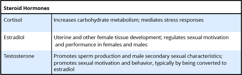
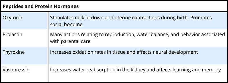
To understand the hormone-behavior relationship, it is important briefly to describe hormones. Hormones are organic chemical messengers produced and released by specialized glands called endocrine glands. Hormones are released from these glands into the blood, where they may travel to act on target structures at some distance from their origin. Hormones are similar in function to neurotransmitters, the chemicals used by the nervous system in coordinating animals’ activities. However, hormones can operate over a greater distance and over a much greater temporal range than neurotransmitters (Focus Topic 1). Examples of hormones that influence behavior include steroid hormones such as testosterone (a common type of androgen), estradiol (a common type of estrogen), progesterone (a common type of progestin), and cortisol (a common type of glucocorticoid) (Table 1, A-B). Several types of protein or peptide (small protein) hormones also influence behavior, including oxytocin, vasopressin, prolactin, and leptin.
Although neural and hormonal communication both rely on chemical signals, several prominent differences exist. Communication in the nervous system is analogous to traveling on a train. You can use the train in your travel plans as long as tracks exist between your proposed origin and destination. Likewise, neural messages can travel only to destinations along existing nerve tracts. Hormonal communication, on the other hand, is like traveling in a car. You can drive to many more destinations than train travel allows because there are many more roads than railroad tracks. Similarly, hormonal messages can travel anywhere in the body via the circulatory system; any cell receiving blood is potentially able to receive a hormonal message.
Neural and hormonal communication differ in other ways as well. To illustrate them, consider the differences between digital and analog technologies. Neural messages are digital, all-or-none events that have rapid onset and offset: neural signals can take place in milliseconds. Accordingly, the nervous system mediates changes in the body that are relatively rapid. For example, the nervous system regulates immediate food intake and directs body movement. In contrast, hormonal messages are analog, graded events that may take seconds, minutes, or even hours to occur. Hormones can mediate long-term processes, such as growth, development, reproduction, and metabolism.
Hormonal and neural messages are both chemical in nature, and they are released and received by cells in a similar manner; however, there are important differences as well. Neurotransmitters, the chemical messengers used by neurons, travel a distance of only 20–30 nanometers (30 X 10–9 m)—to the membrane of the postsynaptic neuron, where they bind with receptors. Hormones enter the circulatory system and may travel from 1 millimeter to >2 meters before arriving at a target cell, where they bind with specific receptors.
Another distinction between neural and hormonal communication is the degree of voluntary control that can be exerted over their functioning. In general, there is more voluntary control of neural than of hormonal signals. It is virtually impossible to will a change in your thyroid hormone levels, for example, whereas moving your limbs on command is easy.
Although these are significant differences, the division between the nervous system and the endocrine system is becoming more blurred as we learn more about how the nervous system regulates hormonal communication. A better understanding of the interface between the endocrine system and the nervous system, called neuroendocrinology, is likely to yield important advances in the future study of the interaction between hormones and behavior.
Hormones coordinate the physiology and behavior of individuals by regulating, integrating, and controlling bodily functions. Over evolutionary time, hormones have often been co-opted by the nervous system to influence behavior to ensure reproductive success. For example, the same hormones, testosterone and estradiol, that cause gamete (egg or sperm) maturation also promote mating behavior. This dual hormonal function ensures that mating behavior occurs when animals have mature gametes available for fertilization. Another example of endocrine regulation of physiological and behavioral function is provided by pregnancy. Estrogens and progesterone concentrations are elevated during pregnancy, and these hormones are often involved in mediating maternal behavior in the mothers.
Not all cells are influenced by each and every hormone. Rather, any given hormone can directly influence only cells that have specific hormone receptors for that particular hormone. Cells that have these specific receptors are called target cells for the hormone. The interaction of a hormone with its receptor begins a series of cellular events that eventually lead to activation of enzymatic pathways or, alternatively, turns on or turns off gene activation that regulates protein synthesis. The newly synthesized proteins may activate or deactivate other genes, causing yet another cascade of cellular events. Importantly, sufficient numbers of appropriate hormone receptors must be available for a specific hormone to produce any effects. For example, testosterone is important for male sexual behavior. If men have too little testosterone, then sexual motivation may be low, and it can be restored by testosterone treatment. However, if men have normal or even elevated levels of testosterone yet display low sexual drive, then it might be possible for a lack of receptors to be the cause and treatment with additional hormones will not be effective.
The endocrine system consists of a series of glands that produce chemical substances known as hormones (Figure 3.30). Like neurotransmitters, hormones are chemical messengers that must bind to a receptor in order to send their signal. However, unlike neurotransmitters, which are released in close proximity to cells with their receptors, hormones are secreted into the bloodstream and travel throughout the body, affecting any cells that contain receptors for them. Thus, whereas neurotransmitters’ effects are localized, the effects of hormones are widespread. Also, hormones are slower to take effect, and tend to be longer-lasting.
Hormones are involved in regulating all sorts of bodily functions, and they are ultimately controlled through interactions between the hypothalamus (in the central nervous system) and the pituitary gland (in the endocrine system). Imbalances in hormones are related to a number of disorders. This section explores some of the major glands that make up the endocrine system and the hormones secreted by these glands (Table 3.2).
Major Glands
The pituitary gland descends from the hypothalamus at the base of the brain and acts in close association with it. The pituitary is often referred to as the “master gland” because its messenger hormones control all the other glands in the endocrine system, although it mostly carries out instructions from the hypothalamus. In addition to messenger hormones, the pituitary also secretes growth hormone, endorphins for pain relief, and a number of key hormones that regulate fluid levels in the body.
Located in the neck, the thyroid gland releases hormones that regulate growth, metabolism, and appetite. In hyperthyroidism or Grave’s disease, the thyroid secretes too much of the hormone thyroxine, causing agitation, bulging eyes, and weight loss. In hypothyroidism, reduced hormone levels cause sufferers to experience tiredness, and they often complain of feeling cold. Fortunately, thyroid disorders are often treatable with medications that help reestablish a balance in the hormones secreted by the thyroid.
The adrenal glands sit atop our kidneys and secrete hormones involved in the stress response, such as epinephrine (adrenaline) and norepinephrine (noradrenaline). The pancreas is an internal organ that secretes hormones that regulate blood sugar levels: insulin and glucagon. These pancreatic hormones are essential for maintaining stable levels of blood sugar throughout the day by lowering blood glucose levels (insulin) or raising them (glucagon). People who suffer from diabetes do not produce enough insulin; therefore, they must take medications that stimulate or replace insulin production, and they must closely control the amount of sugars and carbohydrates they consume.
The gonads secrete sexual hormones, which are important in reproduction, and mediate both sexual motivation and behavior. The female gonads are the ovaries; the male gonads are the testes. Ovaries secrete estrogens and progesterone, and the testes secrete androgens, such as testosterone.
| Major Endocrine Glands and Associated Hormone Functions | ||
|---|---|---|
| Endocrine Gland | Associated Hormones | Function |
| Hypothalamus | Releasing and inhibiting hormones, such as oxytocin | Regulate hormone release from pituitary gland |
| Pituitary | Growth hormone, releasing and inhibiting hormones (such as thyroid stimulating hormone) | Regulate growth, regulate hormone release |
| Thyroid | Thyroxine, triiodothyronine | Regulate metabolism and appetite |
| Pineal | Melatonin | Regulate some biological rhythms such as sleep cycles |
| Adrenal | Epinephrine, norepinephrine | Stress response, increase metabolic activities |
| Pancreas | Insulin, glucagon | Regulate blood sugar levels |
| Ovaries | Estrogen, progesterone | Mediate sexual motivation and behavior, reproduction |
| Testes | Androgens, such as testosterone | Mediate sexual motivation and behavior, reproduction |
Additional Supplemental Resources
Websites
- Areas and Function of the Brain
- Students will interact with the map and chart to review major areas of the brain and their functions. Toggle down on the top left menu to choose different structures to explore.
- Explore the UCLA Laboratory of Neuro Imaging
- We’ve built a diverse team of neurobiologists, mathematicians, and computer scientists, and a worldwide network of collaborators sharing data. Our goal is to increase the pace of discovery in neuroscience by better understanding how the brain works when it’s healthy and what goes wrong in disease.
- Brain Museum
- This web site provides browsers with images and information from one of the world’s largest collection of well-preserved, sectioned and stained brains of mammals. Viewers can see and download photographs of brains of over 100 different species of mammals (including humans) representing over 20 Mammalian Orders.
Videos
- Self Reflected
- Neuroscientist and artist Greg Dunn have created a map of the brain’s neural pathways and animated the firing of neurons. After you have learned about brain regions and neurons, this will provide a beautiful capstone to what you have learned. Closed captioning not available.
- 3 Clues to Understanding Your Brain
- Vilayanur Ramachandran tells us what brain damage can reveal about the connection between cerebral tissue and the mind, using three startling delusions as examples.
- Crossing the Divide: How Neurons Talk to One Another
- View the process of neurotransmission up close in this short video clip. Closed captioning available.
- In-Depth Fight or Flight Response
- This in-depth video explains the cellular processes that result in common “fight or flight” physiological responses, including hair-raising, sweating, and increased respiration. Includes play-by-play printout but not a full transcript.
- Severed Corpus Callosum
- Alan Alda interviews Michael Gazzaniga and a split-brain patient to determine the peculiarities of having a severed corpus callosum. Closed captioning available.
- What is a Neuron?
- This video includes information on topics such as the structure of a neuron. It is hosted by neuroscientist Alie Caldwell. Check out her other videos in the series as well. Closed captioning available.
-
Modern ways of studying the brain |Khan Academy
- This video gives an overview of some of the most common brain imaging tests including CAT, MRI, fMRI, MEG, EEG, and PET.
- Navy SEALs Mental Training
- Video segment from “The Brain: Mystery Explained” documentary featured on The History Channel. Navy SEALs Mental Training: – Goal Setting – Mental Rehearsal (aka Visualization) – Self Talk – Arousal Control
- Crash Course Video #3 – The Chemical Mind
- This video on the chemical mind covers the structure and function of the neuron, neurotransmitters and the endocrine system. Closed captioning available.
- Crash Course Video #4 – Meet Your Master: Getting to Know Your Brain
- This video on getting to know your brain contains information on the structure and function of the cerebral cortex, the limbic system, and lower-level structures. Closed captioning available.
- Anatomy of the Nervous System
- Video on the anatomy of the nervous system
- How the Human Brain Works
- To look at the functions of the brain and neurons, watch this video.
