
PARASITES-NEMATODA
LEARNING OBJECTIVES
Identify representative nematode pathogens
Describe life cycles and unique characteristics of representative roundworms
MCCCD OFFICIAL COURSE COMPETENCIES
Identify structural characteristics of the major groups of microorganisms
Compare and contrast prokaryotic cell and eukaryotic cells
Compare and contrast the physiology and biochemistry of the various groups of microorganisms
Describe the symptoms, associated pathogen, transmission, course, treatment and prophylaxis of common infectious diseases
Apply various laboratory techniques to identify types of microorganisms
MATERIALS
Microscope Demonstrations of:
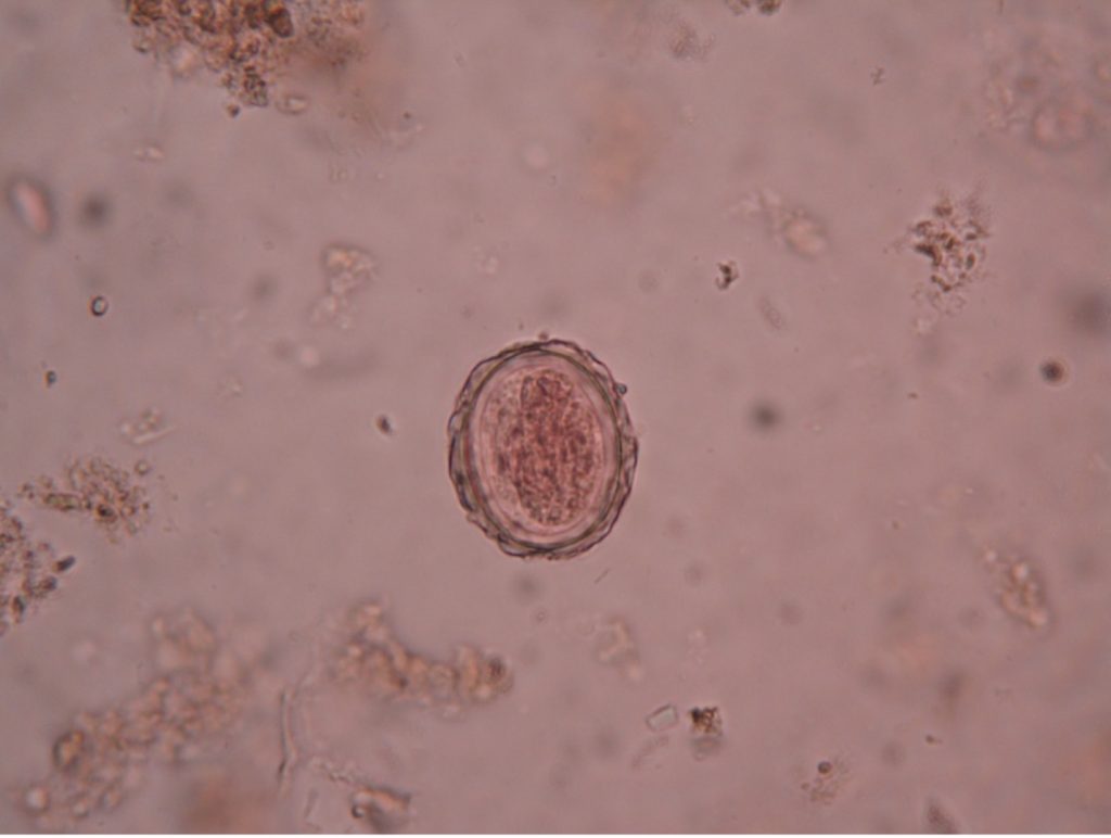
Ascaris lumbricoides ova – 400X total magnification
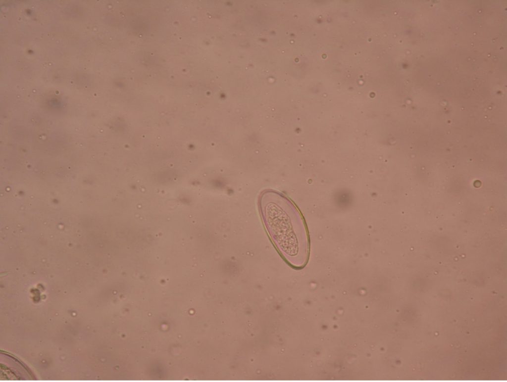
Enterobius vermicularis ova – 400X total magnification
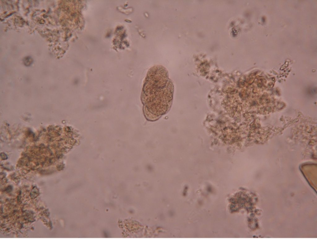
Necator americanus ova – 400X total magnification
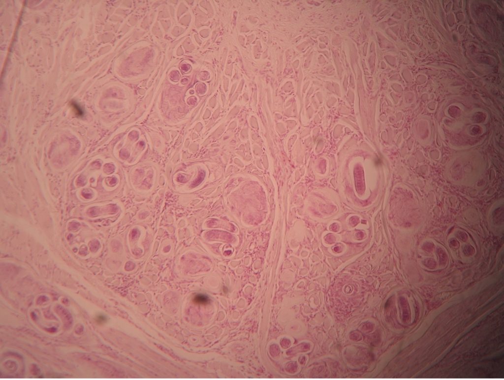
Trichinella spiralis encysted in skeletal muscle – 100X total magnification
PARASITE ALBUM LINK
Symbiosis is an interaction between two organisms living together in a long-term relationship. Symbiotic relationships are classified into three types based on the effect of the interaction on the two organisms. If a one organism benefits from the relationship but the other organism is relatively unaffected, it is classified as commensalism. If both organisms benefit from the symbiotic relationship, it is termed mutualism. If one organism benefits at the expense of the other organism, parasitism occurs. We will examine representative human parasites in three phyla: Nematoda, Platyhelminthes and Protozoa.
Nematodes are often called roundworms because they are cylindrically shaped, resembling a non-segmented earthworm. They have smooth bodies that taper to points at opposite ends. Most roundworms are very small, not exceeding a length of 5 cm.
Most nematodes are harmless and freely inhabit the soil as an essential part of our ecosystem feeding on fungi and bacteria. Some feed on decaying material and thus assist the decomposition process. Most roundworms that parasitize man require a stage outside of the human host to develop into an infective form.
Ascaris lumbricoides (as’kar-is/lum-bri-koy’deez)
The adult Ascaris lumbricoides is rather large, earning the common name “giant intestinal roundworm”. The females, which may be as thick as a pencil, measure 20 to 35 cm (8-12 inches) in length and are larger than the males of the species. Males usually possess a hook-shaped tail helping to distinguish it from the female. The adult Ascaris can live up to 2 years in the small intestines of humans and domestic animals such as pigs and horses. Since only longitudinal muscles are present in the body wall, these worms move only by side-to-side thrashing movements.
The Ascaris ova are covered with a tough cuticle layer that protects the parasite from the gastric juices, enzymes and acid pH of the intestinal tract of the host. After the ovum is swallowed, the larvae hatch and burrow through the intestinal wall to reach the venules or lymphatics. They circulate to the liver, heart and lungs. In the lungs, molting takes place and after ten days the larvae travel up the windpipe to the throat to be swallowed. Ascaris larvae grow to adulthood in the lumen of the small intestine absorbing predigested nutrients from the host’s food supply. Mating occurs in the host’s intestine and the ova are passed from the host in the feces. Both fertile and infertile ova may be seen in Ascaris infections. The fertile egg has a thick, transparent, hyaline shell covered with a thick outer layer. The infertile egg is longer and narrower (90 by 45m) than the fertile egg (60 by 42m) and has a thinner shell. The ova must mature in the soil for two or more weeks before reaching the infective stage for man, and it can remain infective for several months. Because of this maturation cycle in soil, these organisms develop only in warm moist climates.
Ascariasis is a very common roundworm worldwide, but rare in the U.S. Infection occurs mostly in the tropical and subtropical regions with inadequate sanitation. The CDC estimates that 807 million-1.2 billion people are infected with Ascaris lumbricoides worldwide. An individual may harbor as many as one thousand Ascaris worms. The number of worms inside an individual is called the worm burden. A single female Ascaris has a productive capacity of twenty-six million eggs and an average daily output of 200,000 eggs! Ascaris infection is diagnosed by finding ova in the feces.
Small numbers of infecting larvae will produce no obvious symptoms and the first sign of infection may be passage of a live adult worm. Larger numbers may cause transient hepatitis, abdominal pain, intestinal blockage, vomiting, diarrhea, and/or fever. The migrating larvae in the lungs may cause severe pneumonitis, coughing spasms, allergic symptoms and asthmatic breathing. Both pyrantel pamoate and mebendazole have proven to be safe, effective treatments for ascariasis. Proper sanitation and disposal of human feces will control this parasite since the infection is acquired by the fecal-oral route.
Necator americanus (ne-kay’tur/ah-mer-ri-kay’nus)
Necator americanus is a roundworm given the common name “hookworm” describing the curved end of an adult worm. Hookworm ova are passed in the feces already in an early cleavage stage and soon hatch into a larva stage in the soil. The larvae feed on the bacteria and other organic matter in the soil for about a week, then molt into an infective stage. These infective larvae penetrate the intact skin of a bare foot when contact is made with infected soil. The larvae enter the circulation and pass through the right side of the heart and lungs. They are coughed up and swallowed and then reside in the small intestine as adult males and females 1.5 cm (1/2 inch) in length. The adult female can live a few years to 15 years and produce several thousand fertile ova daily. Each ovum has bluntly rounded ends and a single, thin, transparent, hyaline shell.
It is important that the ova of hookworms be identified because of the severe damage they may cause. The adult firmly attaches to the mucosa of the small intestine with its cutting mouth parts and derives its nutrition from the blood of the host. It is estimated that each adult hookworm leaches about 0.15 ml of blood each day; thus, a heavy infection of 500 worms would cause the host to bleed the equivalent of one unit of blood per week. This is sufficient to cause severe anemia and lethargy. It is thought that approximately 500 million persons in the world are infected with hookworm. It is prevalent in rural areas that lack indoor plumbing, proper sanitation, and where the people often choose to go barefoot. Hookworm infestation was common in the southeastern United States, partly due to the moist warm climate there. Improvement in living conditions has greatly reduced hookworm infections.
While hookworm responds to treatment with mebendazole, the control of hookworm relies on proper sanitation and education. In rural communities lacking indoor plumbing, defecation should be limited to specifically constructed pit toilets. Wearing shoes while outdoors would also decrease the transmission of these larvae. Education of the public with regard to the mode of hookworm transmission is essential.
Enterobius vermicularis (en’tur-o’bee-us/vur-mick-yoo-lair’is)
The pinworm, Enterobius vermicularis, is thought to be the most common helminth parasite in temperate regions with a high level of sanitation. While children are more commonly infected than adults, pinworm infestations are found in all ages and socioeconomic groups. Humans are the only host of Enterobius vermicularis.
The life cycle of Enterobius vermicularis is 4-6 weeks. Adults inhabit the cecum and adjacent portions of the large and small intestine of the host. The male pinworm is inconspicuous, about 2 to 5 mm. in length, and no more than 0.2 mm. in width. The female may reach a length of 13 mm. (1/2 inch) and a maximum width of 0.5 mm. The female is distinguished by a long, thin, sharply pointed tail, which gives rise to the common name “pinworm”. The gravid females, containing 11,000 to 15,000 eggs, migrate down the intestinal tract to the anus where they deposit their eggs. Enterobius ova, having a sticky albumin coating, adhere to the perianal region. The eggs mature quickly and are infective within a few hours after they are deposited.
Recovery of eggs from the perianal region provides the most efficient method of diagnosis of pinworm infection. Repeated examinations on consecutive days are necessary because of the irregular migrations of the gravid female worms. Since pinworm eggs are generally deposited at night, the examination should be made in the early morning hours before the patient has washed or defecated. Typically, Graham’s Scotch Tape Method is employed. The sticky side of cellophane tape is applied to the anal area of the patient. The tape is then pressed firmly on a microscope slide, sticky side down. The slide is examined under the low power of a microscope for the presence of pinworm eggs. The eggs are identified by their flattened oval (asymmetrical) shape and possibly the presence of a well-developed embryo.
Migration of the female worm may cause pruritus ani (anal itching). The local itching may cause insomnia and restlessness in children or adults who are infected, as the worms migrate from the anus during the resting hours. In about one-third of infected children, eggs may be obtained from beneath the fingernails. Reinfection of the patient by hand-to-mouth transmission (from scratching the perianal area or handling contaminated fomites) is common and makes control of the parasite difficult. Enterobius ova can survive for 2-3 weeks outside the body in high humidity and moderate temperatures. If towels or linens are shared, these eggs can be passed to others. It is recommended that all members of the infected household be treated simultaneously. Infection of others through contaminated bedding, clothing, bath tubs, toilet seats, dry dust, and air-borne eggs may serve to infect persons at some distance. The prescription drug, mebendazole, is used to treat pinworm. There are over-the-counter drugs available for treatment of pinworms also. Vermox, antiminth, and Povan (pyrvinium pamoate) are acceptable treatments. Patients using Povan should be forewarned that it colors the stool a bright red color!
Trichinella spiralis (trick’i-nell’uh/spy-ray’lis)
Trichinella spiralis is a parasite of carnivorous animals. Bears as well as pigs that are fed uncooked garbage and slaughterhouse scraps can be heavily infected. Infection is initiated by the consumption of raw or insufficiently cooked pork or bear meat containing the encysted larvae of Trichinella spiralis. One pink pork chop can contain 10,000 larvae!
When the encysted larvae pass through the digestive system of the host, the larvae develop into adult worms. Mating of male and female worms occurs, and larvae are produced. The larvae enter the lymphatic vessels where they are transported throughout the body. They leave the capillaries and encyst in the striated muscle, settling in a spiral arrangement. Therefore, Trichinella spiralis is called a tissue parasite.
The symptoms of trichinosis occur 2-8 weeks after ingestion and depend on the number of worms, the size and age of the patient, the tissues invaded, and the general resistance of the patient. While frequently a subclinical infection, trichinosis may lead to death from myocarditis, encephalitis or pneumonitis. Corticosteroids are sometimes beneficial in treating severely symptomatic infections.
Trichinella was once very common in European countries where uncooked garbage containing pork scraps was fed to hogs. Enlightened sanitary practices have resulted in a marked lowering of the rate of infection. In the 1930’s it was estimated that one out of every six persons in the United States became infected with Trichinella spiralis sometime during their life. Approximately 20 cases/year in the last 20 years have been reported in the United States. This decrease is a tribute to the meat inspection program and the stringent laws that have been enacted against feeding uncooked garbage to pigs.
The most common cases of trichinosis are due to homemade jerky and sausage. Smoking, salting, or drying the meat does not destroy the infective larval forms, but prolonged freezing (20 days in the average home freezer) will decontaminate pork. Special care must be taken with large pork products to ensure that the internal temperature of the meat reaches a minimum of 165 degrees Fahrenheit.
Dracunculus medinensis (druh-kunk’u-lus/med-eh-nin’sus)
This so-called “guinea worm” used to be found in Asia, Africa, and the Middle East. Due to a massive initiative led by former U.S. president, Jimmy Carter, the number of cases worldwide has dropped from 3.5 million in 1986 to fewer than 30 cases currently. The parasite has been eradicated from most of the world and is now found primarily in Ghana and the Sudan.
People become infected by drinking contaminated water that carry water fleas infected with the larvae of the parasite. The larvae are released in the peritoneal space. After the males and females mate, the male dies. The female, and the larvae she produces, need to escape their human host. The female forms a fiery blister somewhere in the body and eventually works her way to freedom. This can take many weeks – sometimes as long as three months. To make matters worse, the female is about 36 inches long. People used to try to pull the worm out but found the worm would break and the wound would become infected. So, infected hosts learn to wind the worm around a stick and attach the entire apparatus to their bodies, slowly winding the worm around the stick as it leaves the body. The blister made by the worm is very painful and people cool the blister in water–water also used for drinking. This causes the blister to break open, releasing the larvae of the worm to infect water fleas which begins the cycle again.
Education about the life cycle of the parasite, sterile medical supplies, and filters that can be used to filter drinking water have resulted in the containment of this parasite and possible eradication from the earth.
Zoonotic infections
Toxocara (tox-o-care’-a)
Toxocara canis (dogs) and Toxocara cati (cats) are responsible for zoonotic infections in humans. Most cases are not serious, and many people do not notice symptoms from these small worms. The most severe cases are usually found in small children. Puppies usually get infected from their mother before birth or from her milk. The puppies expel large numbers of eggs in their feces when they are about a month old. Children get infected from eating dirt or playing in dirt that contain feces from infected dogs and cats and then putting their hands in their mouths. People who demonstrate symptoms usually have ocular larva migrans (OLM), and infection of the eye that causes scarring of the retina. Others demonstrate inflammation from the movement of the worms through the body. This is called visceral larva migrans or VLM.
Dirofilaria immitis (dye’ro-fill-air’-ee-uh/im’-i-tus)
Some roundworms are very tiny and transmitted by an insect vector. An example is Dirofilaria immitis that infects dogs, cats, coyotes, and wolves in very warm or tropical areas. The mosquito is the vector. The female mosquito injects larvae into the host that develop into male and female microfilaria in the blood stream. These worms wind up in the heart and lungs of these animals. They rarely are transmitted to humans.
PRE-ASSESSMENT
procedure
Complete the Nematoda portion of the parasite lab practical study guide.
parasite practical practice
POST TEST
DISCOVERIES IN MICROBIOLOGY
DR. SYDNEY BRENNER
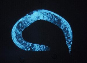 South African biologist Dr. Brenner turned the Caenorhabditis elegans nematode worm into a model system for human disease research in the 1960’s and 1970’s. Brenner chose the worm because it was more complex than other well understood organisms, such as bacteria, yet still simple enough to study in depth. C. elegans is still widely used in biology today. The research database PubMed lists some 15,000 papers published in the past decade that include a reference to the worm. Brenner also co-discovered messenger RNA. These intermediary molecules convey a cell’s genetic code, which is written in DNA, to the cellular machinery that translates messenger RNA into a protein. And, alongside Francis Crick and others, he worked out that the genetic code of DNA is made up of a series of triplets of nucleotides called codons, which encode the amino acids that make up a particular protein.
