
VIRAL PLAQUE ASSAY
LEARNING OBJECTIVES
Discuss viral growth requirements
Demonstrate and recognize viral plaques
Perform standard dilutions
Calculate the number of organisms in a diluted sample
MCCCD OFFICIAL COURSE COMPETENCIES
Compare and contrast viruses and cells
Describe the modes of bacterial and viral reproduction and proliferation
Utilize aseptic technique for safe handling of microorganisms
Apply various laboratory techniques to identify types of microorganisms
Identify structural characteristics of the major groups of microorganisms
Compare and contrast prokaryotic cell and eukaryotic cell
Compare and contrast the physiology and biochemistry of the various groups of microorganisms
MATERIALS
Students will complete the lab in groups of 3 at Pecos and groups of 4 at Williams
Media:
2 tubes each containing 9.9 ml Luria broth
3 tubes each containing 9.0 ml Luria broth
5 tubes of 5 ml of melted agar in 50oC degree water bath (top agar)
5 Luria Agar plates
Equipment:
5 small sterile microcentrifugue tubes
10 sterile transfer pipettes
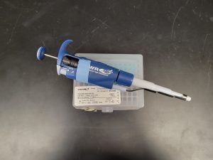
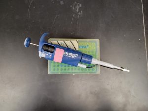
Automatic pipettors- 0.1ml (pink tape pipette) and 1.0ml (blue tape pipette)
Box of sterile pipette tips
Stock Cultures:
Escherichia coli B strain broth culture 8/2 lab sections at Pecos 6/2 lab sections at Williams
T4 bacteriophage broth culture 8/2 lab sections at Pecos 6/2 lab sections at Williams
BACTERIA ALBUM LINK
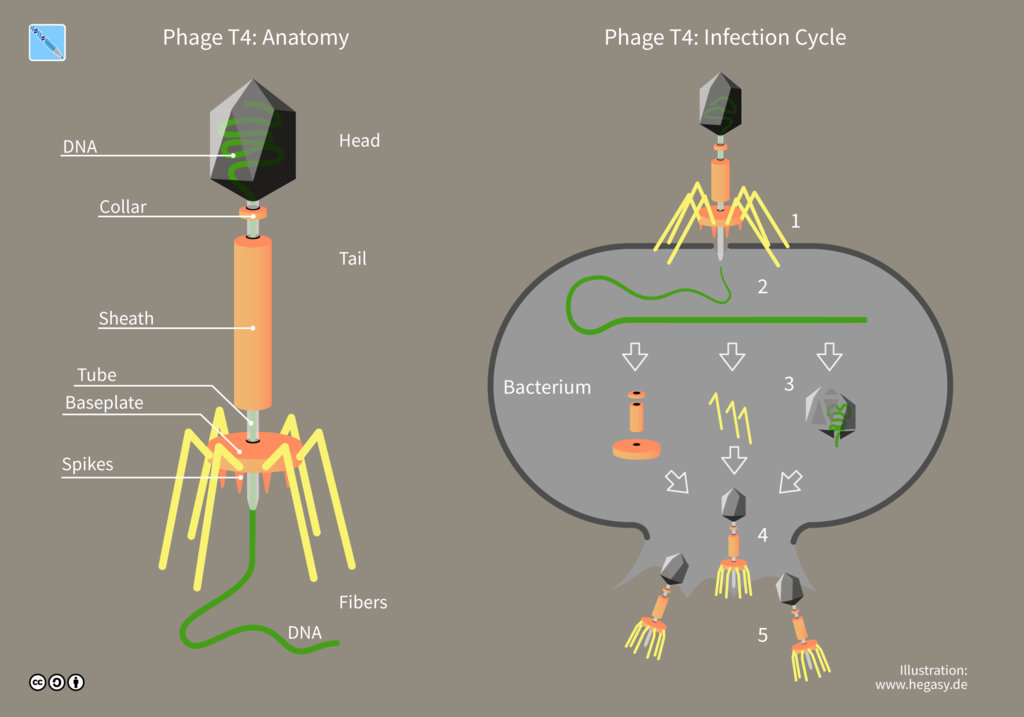
Viruses are obligate intracellular parasites, meaning they can only replicate inside another living host cell. Therefore, ordinary agar or broth media suitable for growing bacterial cultures will not sustain viral growth. However, bacteria growing in lab media can serve as the host cell for growing viruses that infect bacteria.
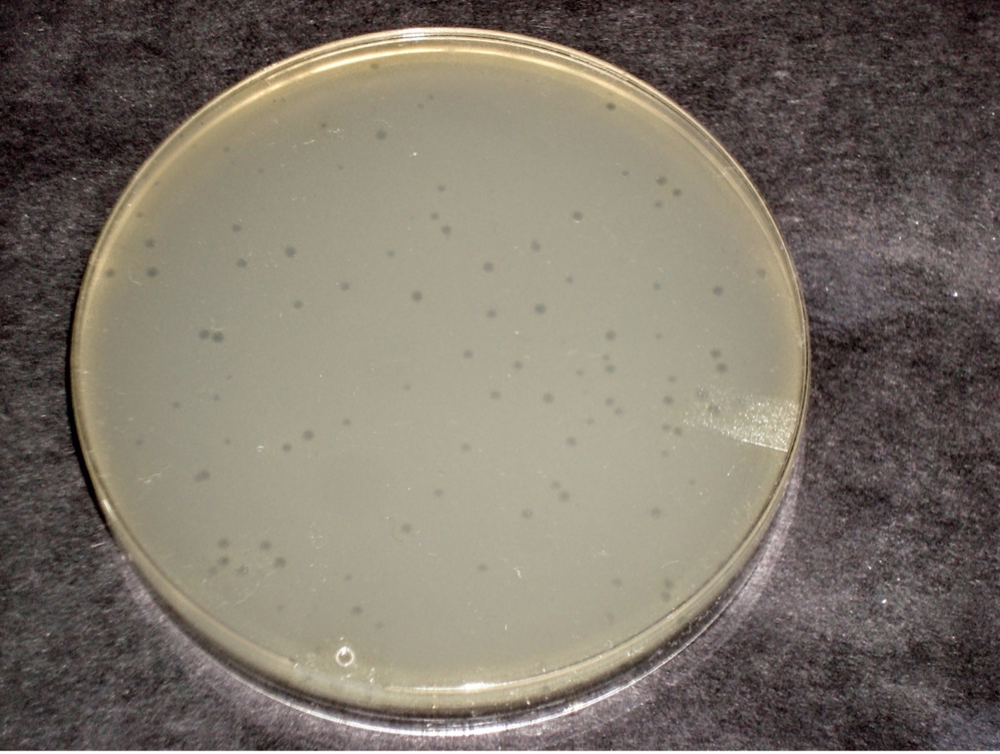
A virus that infects bacteria is called a bacteriophage or phage. A phage parasitizes bacteria and utilizes bacterial cell components to produce progeny virus. Lytic viruses burst open and kill their bacterial host once the appropriate number of viruses has been synthesized. A plaque is an area of clearing in a confluent lawn of bacterial growth which represents the spot where a virus has landed, infected the bacteria it encountered, and lysed them. Each plaque represents a virus particle or plaque-forming unit (PFU). Since there are too many bacteriophage in the original sample, it is best to dilute the sample to obtain a countable plate in which 30-300 individual plaques can be seen. We will be using the T4 bacteriophage and T4 host cells, E. coli strain B.
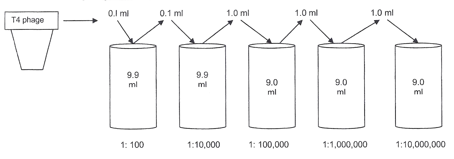
Serial dilution of the T4 phage begins the quantitation procedure.
Diluting the original T4 phage sample:
To calculate the dilution factor used for a sample, compare the amount of sample to the total volume of broth in the container.
0.1 ml of T4 phage sample to be tested
0.1 ml of sample + 9.9 ml of broth = 10 ml total volume in tube = 1:100
The sample added to the next tube is not the original sample. It is the original sample already diluted 1:100. Thus, the previous dilution is multiplied by the next dilution.
0.1 ml of diluted sample
0.1 ml of diluted sample+ 9.9 ml of broth = 1:10,000 dilution of the original T4 phage sample
The sample added to the next broth is taken from the previous dilution. Thus, this sample has already been diluted 10,000 times.
1 ml of the further diluted sample
1 ml of diluted sample + 9.0 ml broth = 1:100,000 dilution of the original T4 phage sample
You will continue diluting the original sample to obtain further dilutions of 1:1,000,000 and 1:10,000,000. These dilutions will help you find a dilution of virus in which 30-300 plaques can be counted.
Calculating the PFU:
The reason for performing the exercise is to count the T4 phages (PFU) in 1.0 ml of the original sample. To determine this, find the plate that has the countable number of PFU and count the plaques. This plaque number must be multiplied by the dilution (used to make that plate) and the amount of the sample taken.
PRE-ASSESSMENT
PROCEDURE
Students will complete the lab in groups of 3 at Pecos and groups of 4 at Williams
Making serial dilutions of T4 Phage
1. Obtain 2 tubes of Luria broth, each containing 9.9 ml. Also obtain 3 tubes of Luria broth, each containing 9.0 ml. Place the tubes in the proper order in a rack as shown in the figure below. Label the tubes as 1:100, 1:10,000, 1:100,000, 1:1,000,000, 1:10,000,000 as shown in the figure below.
FOR EACH OF THE FOLLOWING DILUTIONS: Always use a new pipette tip and transfer pipette. The pipettes volumes are already set for you.
1. Using the pipette with the pink tape and a sterile yellow pipette tip, transfer 0.1 ml bacteriophage into the luria broth tube labeled 1:100. Discard the tip in the biohazard trash. Mix the broth using a transfer pipette by taking in and expelling the fluid back into the tube several times. Discard the transfer pipette in the biohazard trash.
2. Using the pipette with the pink tape and a sterile yellow pipette tip, transfer 0.1 ml of the 1:100 dilution into the luria broth labeled 1:10,000. Mix the broth using a transfer pipette by taking in and expelling the fluid back into the tube several times. Discard the transfer pipette in the biohazard trash.
3. Using the pipette with the blue tape and a sterile blue pipette tip, transfer 1.0 ml of the 1: 10,000 dilution into the luria broth tube labeled 1:100,000. Mix the broth using a transfer pipette by taking in and expelling the fluid back into the tube several times. Discard the transfer pipette in the biohazard trash.
4. Using the pipette with the blue tape and a sterile blue pipette tip, transfer 1.0 ml of the 1: 100,000 dilution into the luria broth tube labeled 1:1,000,000. Mix the broth using a transfer pipette by taking in and expelling the fluid back into the tube several times. Discard the transfer pipette in the biohazard trash.
5. Using the pipette with the blue tape and a sterile blue pipette tip, transfer 1.0 ml of the 1: 1,000,000 dilution into the luria broth tube labeled 1:10,000,000. Mix the broth using a transfer pipette by taking in and expelling the fluid back into the tube several times. Discard the transfer pipette in the biohazard trash.
Mix bacteriophage dilutions with E.coli
1. Label 5 sterile microcentrifuge tubes the same way you labeled the original test tube dilutions – 1:100, 1:10,000, 1:100,000, 1:1,000,000, 1:10,000,000.
2. Pipette 0.1 ml E. coli into each of the microcentrifuge tubes using one pipette tip.
3. Using a different pipette tip for each transfer, pipette 0.1 ml of each phage dilution into the corresponding microcentrifuge tube of E. coli. (0.1 ml of 1:100 phage dilution into the tube of E. coli labeled 1:100, etc.) Rotate the tubes in your palms to mix them.
4. Incubate the microcentrifuge tubes containing the bacteriophage and E. coli at room temperature for 5 minutes.
5. Remove the labels from the luria broth dilutions and place the tubes in the discard rack to be autoclaved.
6. Return the E. coli culture.
Plating the phage/E.coli mixture
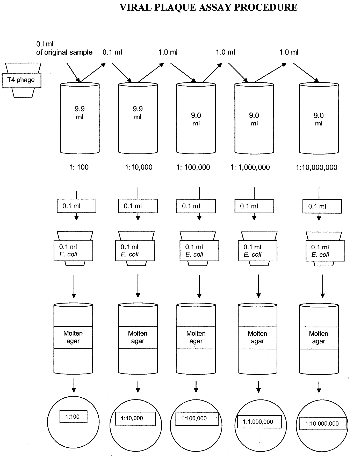
1. Obtain 5 Luria agar plates. Label the lid (not the bottom) of each plate with your group name and the corresponding dilutions of the phage/E.coli mixtures (1:100, 1:10,000, 1:100,000, 1:1,000,000, 1:10,000,000). The plates will be incubated lid side up to prevent the agar lawn from falling off.
2. Obtain one tube of melted top agar from the water bath. (ONLY REMOVE ONE TUBE OF AGAR AT A TIME – IT SOLIDIFIES QUICKLY!).
3. Transfer the contents of the E. coli/phage mixture with a sterile transfer pipette (do not use a pipette and pipette tip) into the melted top agar and gently mix. Immediately pour the contents onto the corresponding Luria agar plate. Quickly swirl the plate gently so that the liquid agar spreads evenly over the surface of Luria agar. Avoid producing bubbles which will affect the results.
4. Repeat Step 2 for each phage/E.coli dilution.
5. Incubate all plates right side up in the class plate tray at 37o C until the next lab session.
6. Dispose of the microcentrifuge tubes in the biohazard trash.
7. Place the tubes that contained the melted luria agar in the discard rack.
Calculating the number of bacteriophages in the original sample
1. Obtain the 5 agar plates and spread them out in order of dilution. Determine which dilution of bacteriophage produced between 30-300 plaques on the agar plate. Dispose of the other dilution plates in the biohazard trash.
2. Count the number of plaques on the agar plate and record the number on the worksheet.
3. Calculate the number of bacteriophage (Plaque Forming Units) in 1 ml of the original sample.
Plaque count x Plate Dilution x 10 (if you plated 0.1 ml)
You need to multiply by 10 if you plate 0.1 ml because we are calculating PFU/ml not PFU/0.1ml
Let’s do an example. You have performed a serial dilution of a sample with an unknown concentration of microorganisms. You counted 122 plaques on a plate inoculated with 0.1 ml of a 1:10,000 dilution. What was the population density of the original sample per milliliter (PFU/ml)?
122 x 10000 x 10 = 12,200,000 PFU/ml
To express 12,200,000 in scientific notation it needs to be a number between 1-9. The decimal point is at the end of the number, move the decimal to the left until you have a number between 1-9. The exponent will be positive.
1.22 x 10⁷ PFU/ml
4. Report the result in scientific notation on the worksheet.
POST TEST
DISCOVERIES IN MICROBIOLOGY
DR. ESTER LEDERBERG
Before 1953, all viruses were considered lytic. In 1953, American microbiologist Dr. Esther Lederberg published her discovery of the lambda bacteriophage and lysogenic replication. Lambda phage is now used ubiquitously in molecular biology to study gene regulation and recombination. Dr. Lederberg also invented replica plating. A petri plate containing bacterial colonies is inverted, and its surface is pressed against a cylindrical block covered with velvet pad. Sterile plates are inverted and pressed against the velvet pad disc to pick up samples of the colonies.
