SIMPLE STAIN
LEARNING OBJECTIVES
Properly perform the simple stain technique
Identify morphology of bacteria
Identify arrangement of bacteria
MCCCD OFFICIAL COURSE COMPETENCIES
Utilize aseptic technique for safe handling of microorganisms
Apply various laboratory techniques to identify types of microorganisms
Identify structural characteristics of the major groups of microorganisms
Compare and contrast prokaryotic cell and eukaryotic cell
Compare and contrast the physiology and biochemistry of the various groups of microorganisms
MATERIALS
Equipment:
“E” slide and “S” slide prepared in the streak plate lab exercise
Bibulous paper
Lens wipes
Stain container
Slide warmer
Microscope
Immersion oil
Stains:
Crystal violet
Safranin
BACTERIA ALBUM
Unstained bacteria are colorless. To see them with the microscope we often use chemical compounds called stains. Staining improves the ability to see small, colorless bacteria. To understand how staining works, it will be helpful to know a little about the physical and chemical nature of stains. Stains are generally salts in which one of the ions is colored. A salt is a compound composed of a positively charged ion and a negatively charged ion. For example, the stain methylene blue is the salt methylene blue chloride which will dissociate in water into a positively charged methylene blue ion which is blue in color and a negatively charged chloride ion which is colorless.
Stains may be divided into two groups: basic and acidic. If the color portion of the stain resides in the positive ion, it is called a basic stain. Some examples of basic stains include methylene blue, crystal violet, and safranin. If the color portion of the stain is in the negative ion, it is called an acidic stain. Some examples of acidic stains include nigrosin, congo red and eosin.
Because of its chemical nature, the cytoplasm of all bacterial cells has a slight negative charge when growing in a medium of near neutral pH. Therefore, when using a basic stain (positively charged), the positively charged color portion of the stain combines with the negatively charged bacterial cytoplasm staining the bacteria.
In a simple stain, the bacteria are stained with one positively charged stain. Crystal violet, methylene blue, and safranin are all examples of positively charged stains used in simple stains, although any positively charged stain may be used.
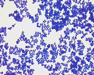
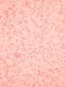
Simple stains are easy to perform and provide basic information about size, morphology (shape), and arrangement of the bacteria. When viewing a simple stain, the microorganisms will appear one color against a clear background.
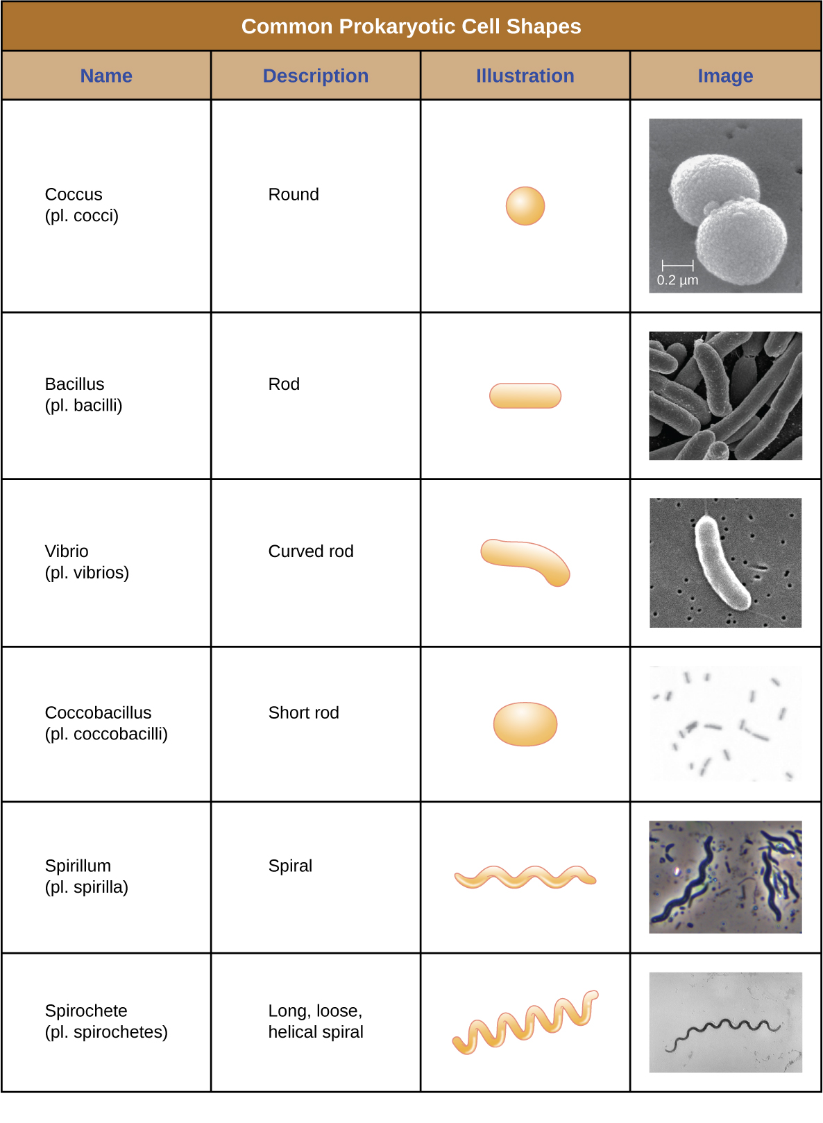
Although thousands of bacteria have been identified, only a handful of cell morphologies (shapes) are commonly seen microscopically. The figure below names and illustrates bacterial morphologies.
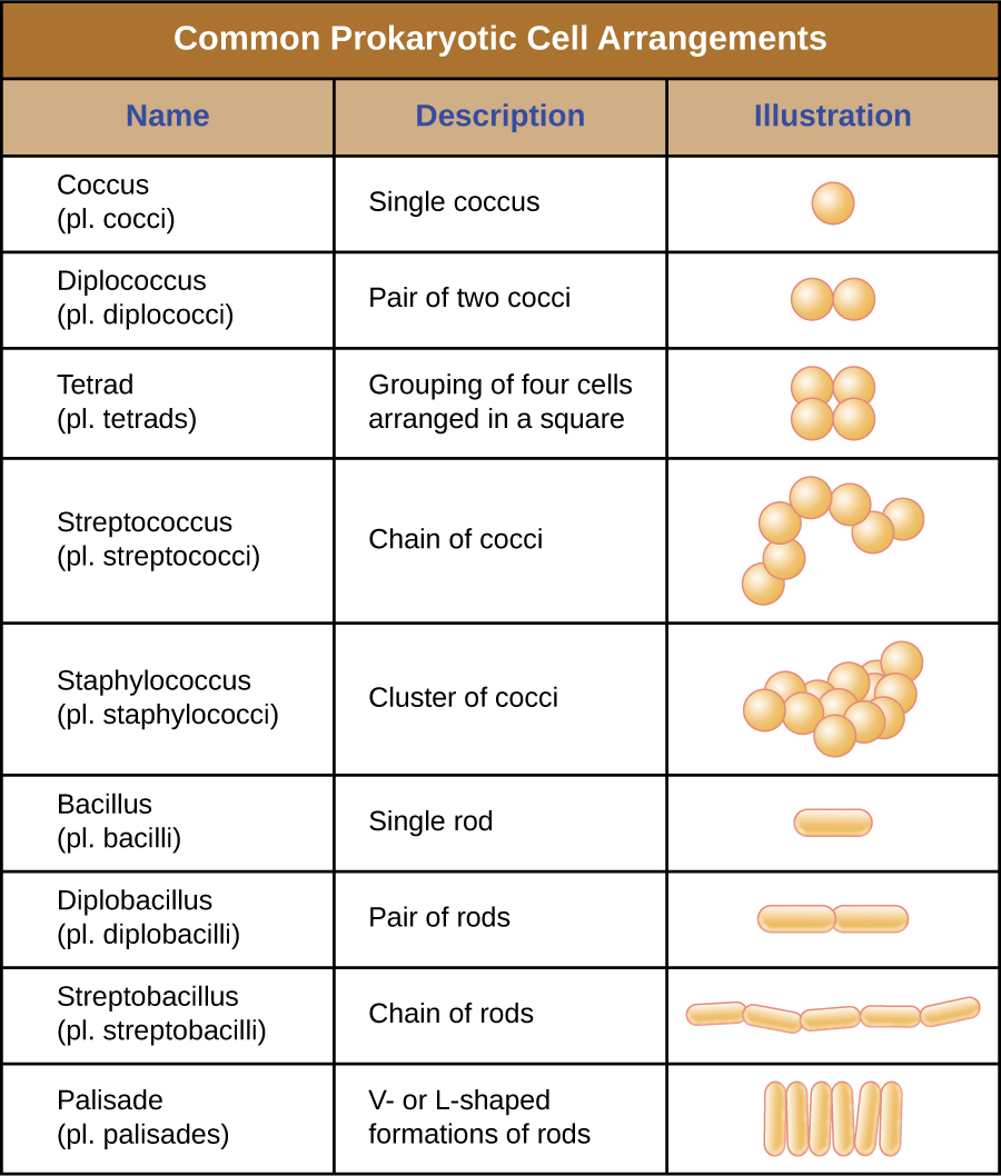
In addition to cellular shape, bacteria may group together in certain distinctive arrangements depending on the plane of cell division. Some common arrangements are shown in the figure below.
PRE-ASSESSMENT
PROCEDURE
Simple stain the “E” and “S” slides you prepared in the streak plate lab exercise.
Step 1. Add a few drops of positively charged crystal violet OR positively charged safranin to the smear. Wait 1 minute. Rinse the smear with deionized water. Shake excess water off the smear.
Step 2. Gently blot the excess water from the slide using bibulous paper. View the stained slides.
Observe stained slides
Step 1. Review how to set up your microscope properly in the microscope lab exercise. Bacteria on the stained smear will need to be magnified 1000X to be able to determine morphology and arrangement.
Step 2. Draw the simple stained bacteria you have observed at 1000X total magnification in the worksheet.
Step 3. After you have observed the simple stain slides, dispose of the slides in the used slide basin.
Post test
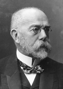
DISCOVERIES IN MICROBIOLOGY
Robert Koch introduced bacterial staining techniques laying the foundation for visualizing bacteria under a microscope. Robert Koch and his team were able to identify the bacterial causes of tuberculosis (1882) and cholera (1883). Adopting Koch’s method, other researchers were able to identify the bacteria that caused diseases such as typhus (1880), tetanus (1884) and the plague (1894).