
IDENTIFYING GRAM POSITIVE BACTERIA DEMONSTRATION LAB
LEARNING OBJECTIVES
Discuss the biochemical tests used to identify Staphylococcus.
Differentiate Staphylococcus from other Gram-positive cocci found in clusters on Gram stain.
MCCCD OFFICIAL COURSE COMPETENCIES
Describe the modes of bacterial and viral reproduction and proliferation.
Apply various laboratory techniques to identify types of microorganisms.
Identify structural characteristics of the major groups of microorganisms.
Compare and contrast prokaryotic cell and eukaryotic cell.
Compare and contrast the physiology and biochemistry of the various groups of microorganisms.
THIS IS A DEMONSTRATION LAB. PLEASE READ THROUGH THE LAB EXERCISE. RECORD THE TEST RESULTS FOR UNKNOWN 1, 2, 3, AND 4 IN THE IDENTIFYING GRAM POSITIVE BACTERIA QUESTION DOCUMENT.
Gram stains and other differential stains can help us group microorganisms, but we can use selective and differential media and biochemical testing to identify these organisms according to their genus and species. In this lab exercise, you will identify four members of the Micrococcaceae family (Staphylococcus aureus, Staphylococcus epidermidis, Micrococcus luteus and Micrococcus roseus). These are normally found on the skin and mucous membranes in the mouth, nasal passages, throat, anus and vagina.
Staphylococcus aureus is usually considered to be pathogenic, but it may be found in the absence of disease. Some people are carriers of this organism, meaning they harbor the pathogen but exhibit no symptoms. One study found as many as 90% of hospital personnel were S. aureus carriers! This poses a risk for hospitalized patients whose immune systems may already be stressed or compromised by their underlying condition. MRSA is especially dangerous because these isolates have become resistant to penicillin derivatives, such as methicillin and oxacillin.
Staphylococcus aureus produces enzymes (hyaluronidase and collagenase) to help it invade and parasitize healthy tissue. Staphylococcus aureus produces coagulase and staphylokinase that hijack the host’s coagulation system. It can cause infections ranging from superficial (boils, impetigo) to systemic infections which involve deeper tissues, such as bone (osteomyelitis), lung (pneumonia), heart valves (endocarditis), brain (meningitis), and blood (toxic shock syndrome). Staphylococcus aureus is the most common etiologic agent of wound infections, including surgical infections. It is a common cause of Healthcare Acquired/Associated Infections (HAI) which are acquired in hospitals and other healthcare facilities. And, many strains produce an exotoxin which causes food poisoning.
Staphylococcus epidermidis is the predominant species of Staphylococcus found on the skin and usually not pathogenic. Micrococcus luteus and Micrococcus roseus are also considered to be avirulent, though it is important to remember that under the proper circumstances, almost any bacteria can cause disease.
Selective and Differential media and biochemical testS overview
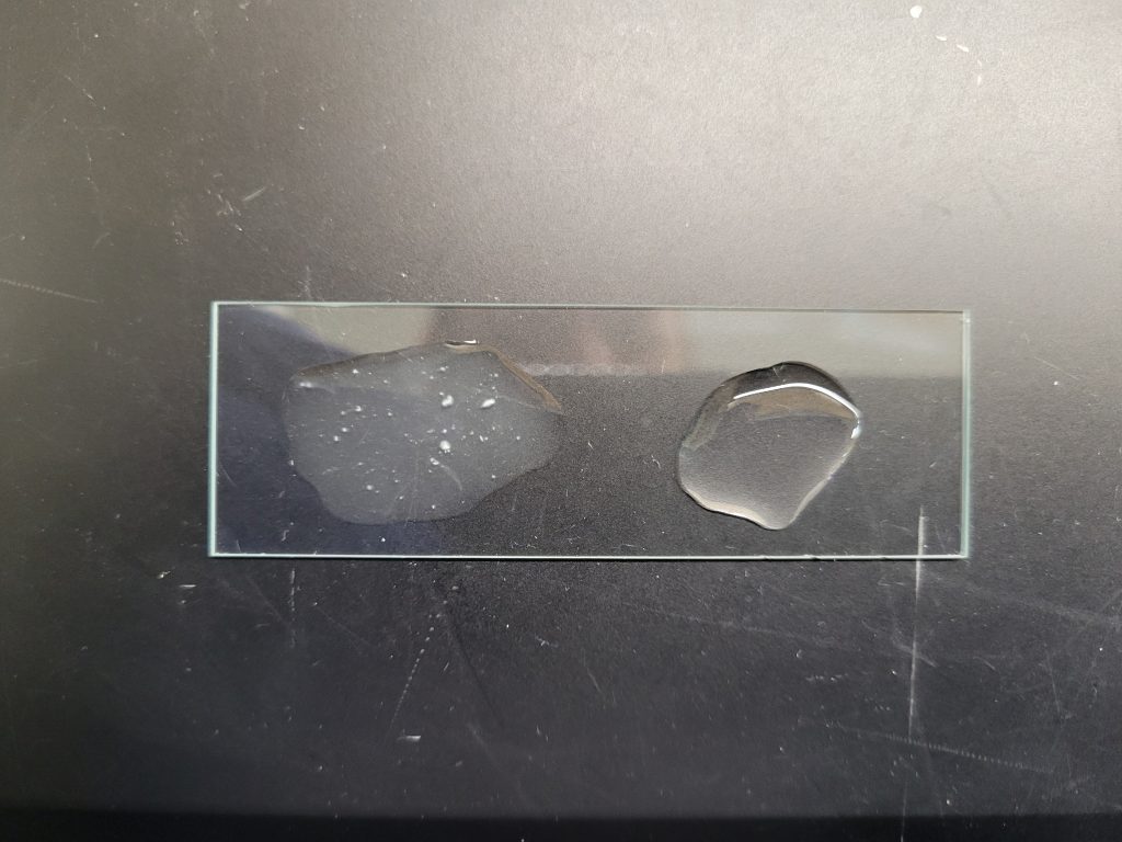
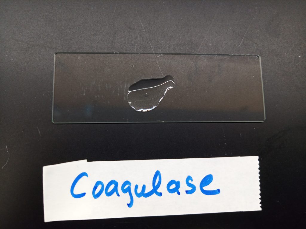
COAGULASE TEST
This biochemical test determines the ability of the bacteria to produce bound coagulase. This exoenzyme will cause the conversion of fibrinogen in plasma to fibrin and form a clot. The fibrin covers the surface of the bacteria, making it resistant to phagocytosis. A large amount of inoculum is mixed with rabbit plasma on a slide. Rabbit plasma provides the fibrinogen. Obvious clotting of the rabbit plasma indicates the organism produces bound coagulase.
Clotting = Positive
Clear = Negative
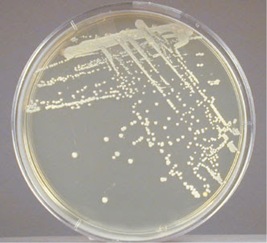
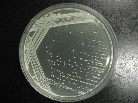
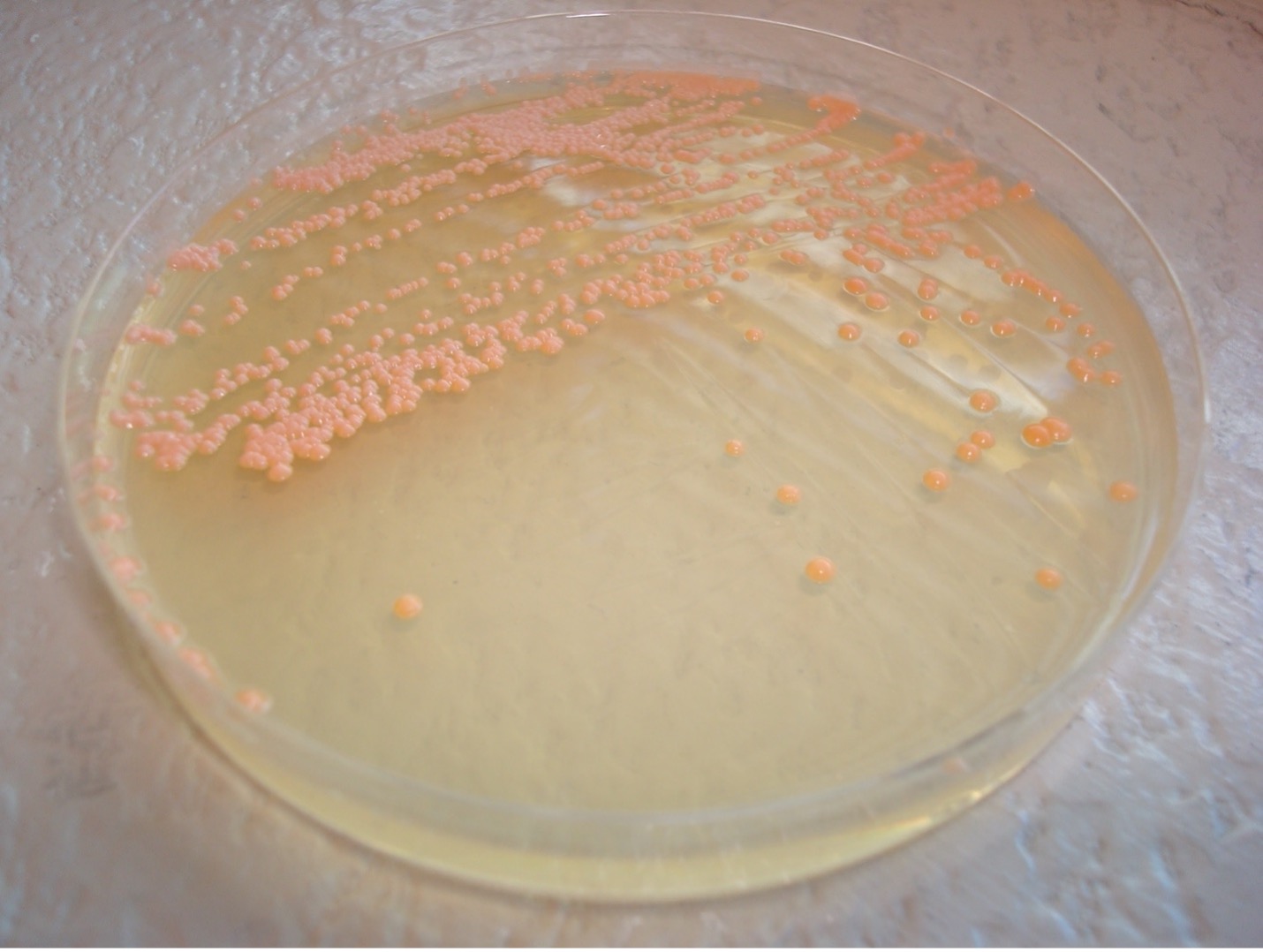
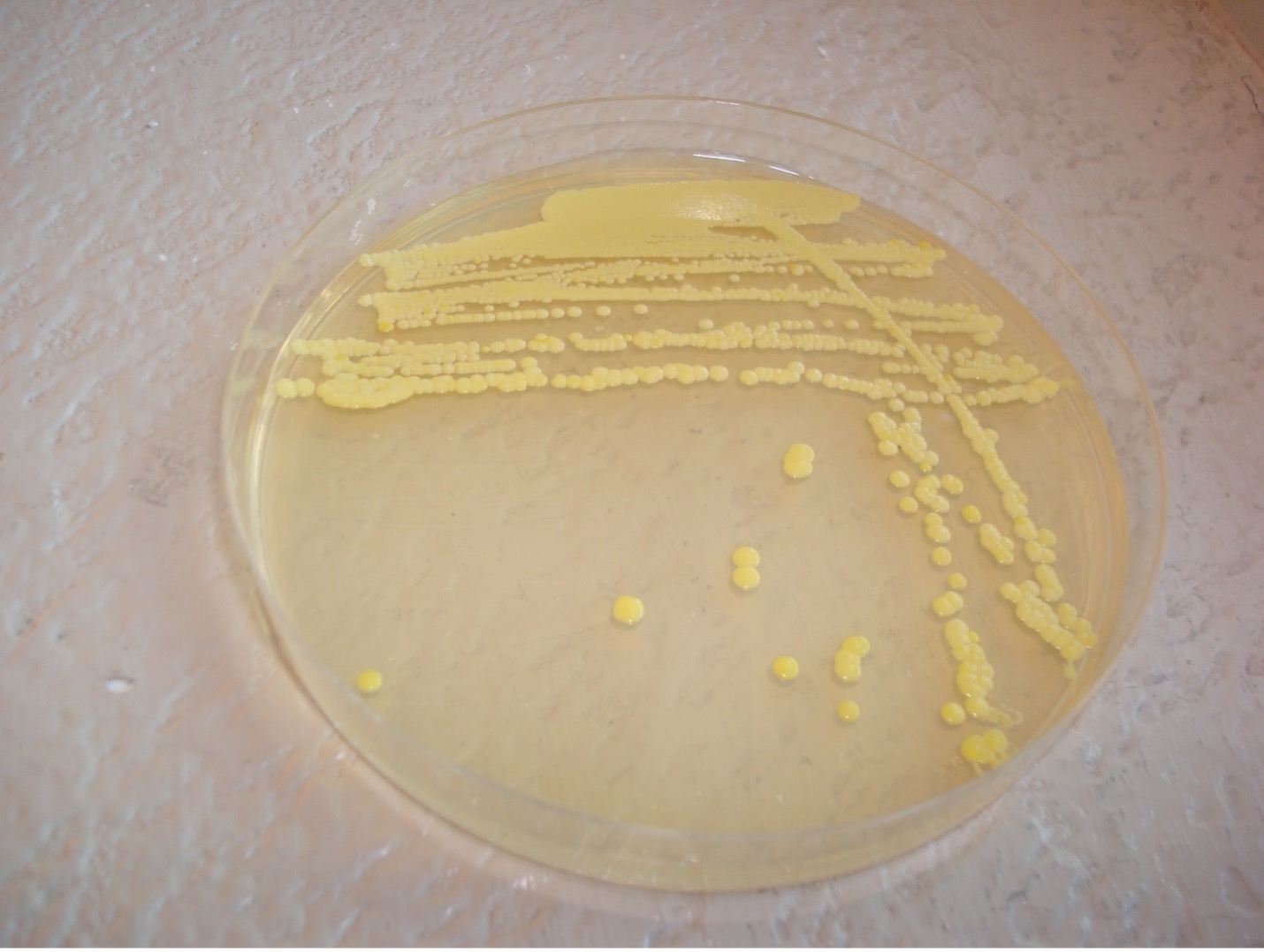
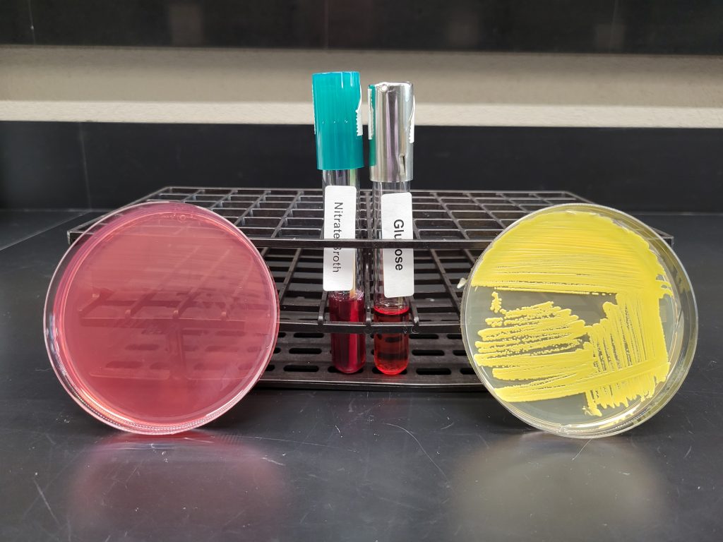
PIGMENT PRODUCTION
Staphylococcus will produce a white pigment while some species of Micrococcus will produce colorful pigment on TSA agar which can serve as an identifying characteristic.
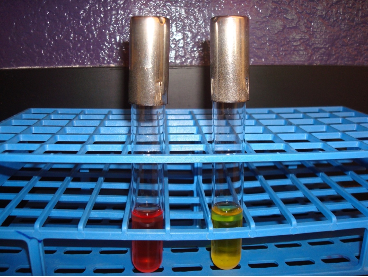
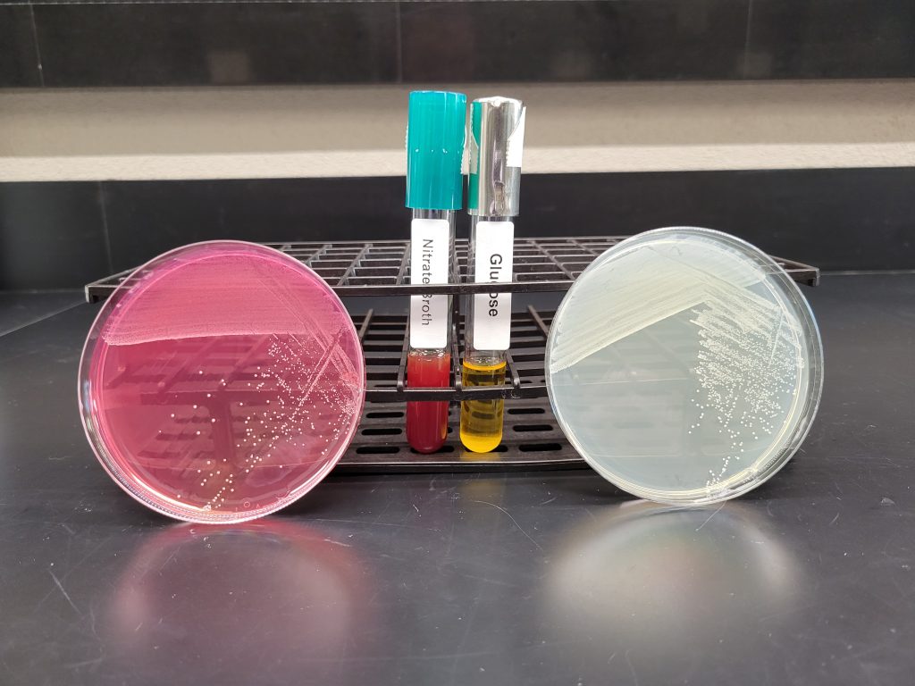
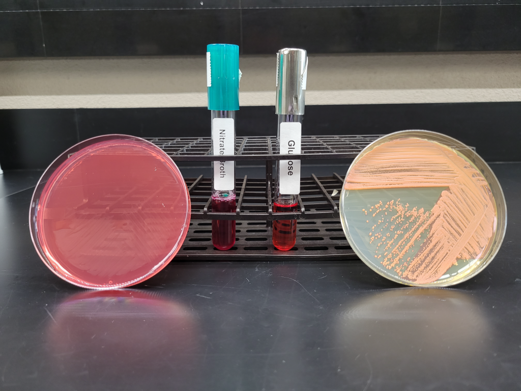
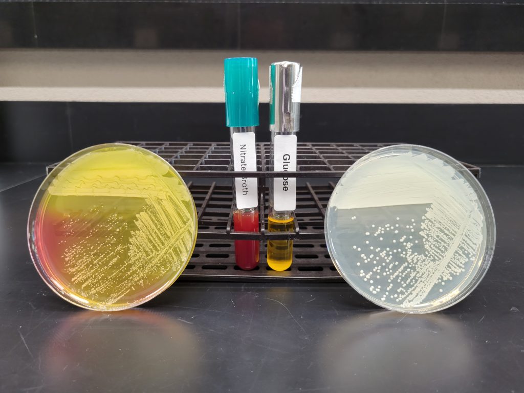
GLUCOSE FERMENTATION TEST
This differential media detects the ability of the bacterium to break down glucose to pyruvic acid. If acid is produced, the pH indicator will turn yellow.
AFTER INCUBATION
Yellow broth = Positive test
Red broth = Negative test
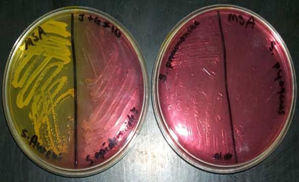
MANNITOL SALT AGAR
This is a selective and differential media which grows and differentiates Staphylococcus species. Since Staphylococcus is salt-tolerant, it will be able to grow in 7.5% salt and Staphylococcus aureus will be differentiated by mannitol fermentation.
AFTER INCUBATION
Evaluate how the well the organism tolerated the salt in the agar.
Bacteria grew very well = Positive
Bacteria were inhibited from growing = Negative
If the bacteria grew very well on the media, then evaluate the organism’s ability to ferment the mannitol in the agar.
Media turned yellow = Positive = mannitol was fermented (Do not confuse the yellow pigmented colonies of Micrococcus luteus as mannitol fermentation)
Media stayed reddish pink = Negative = mannitol was NOT fermented
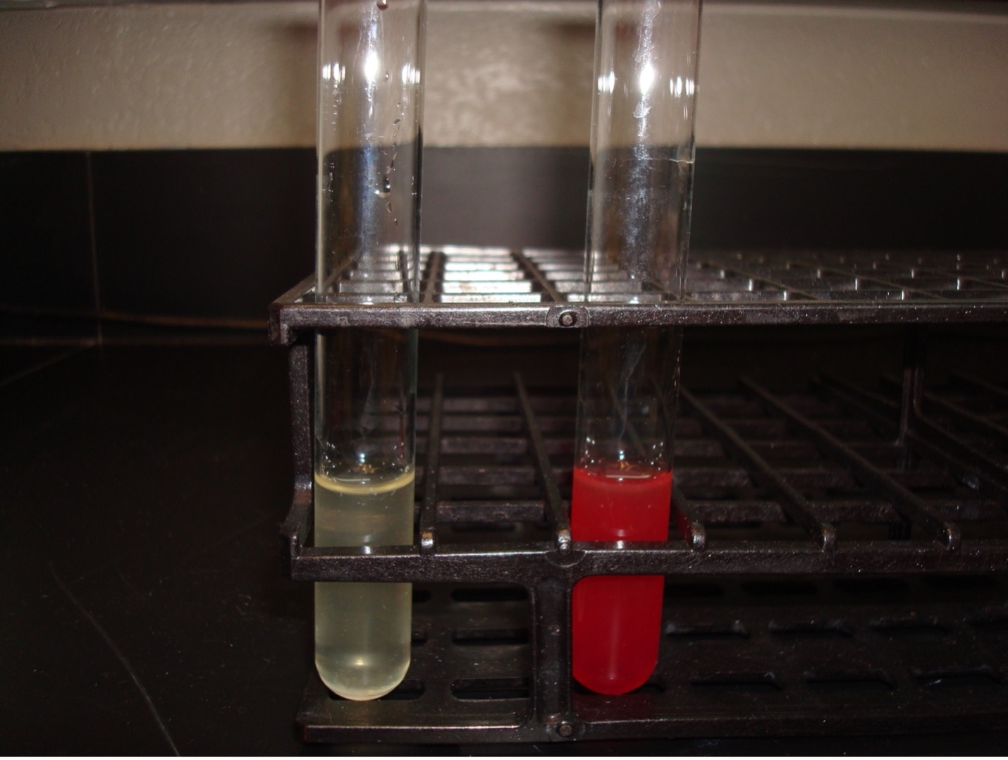
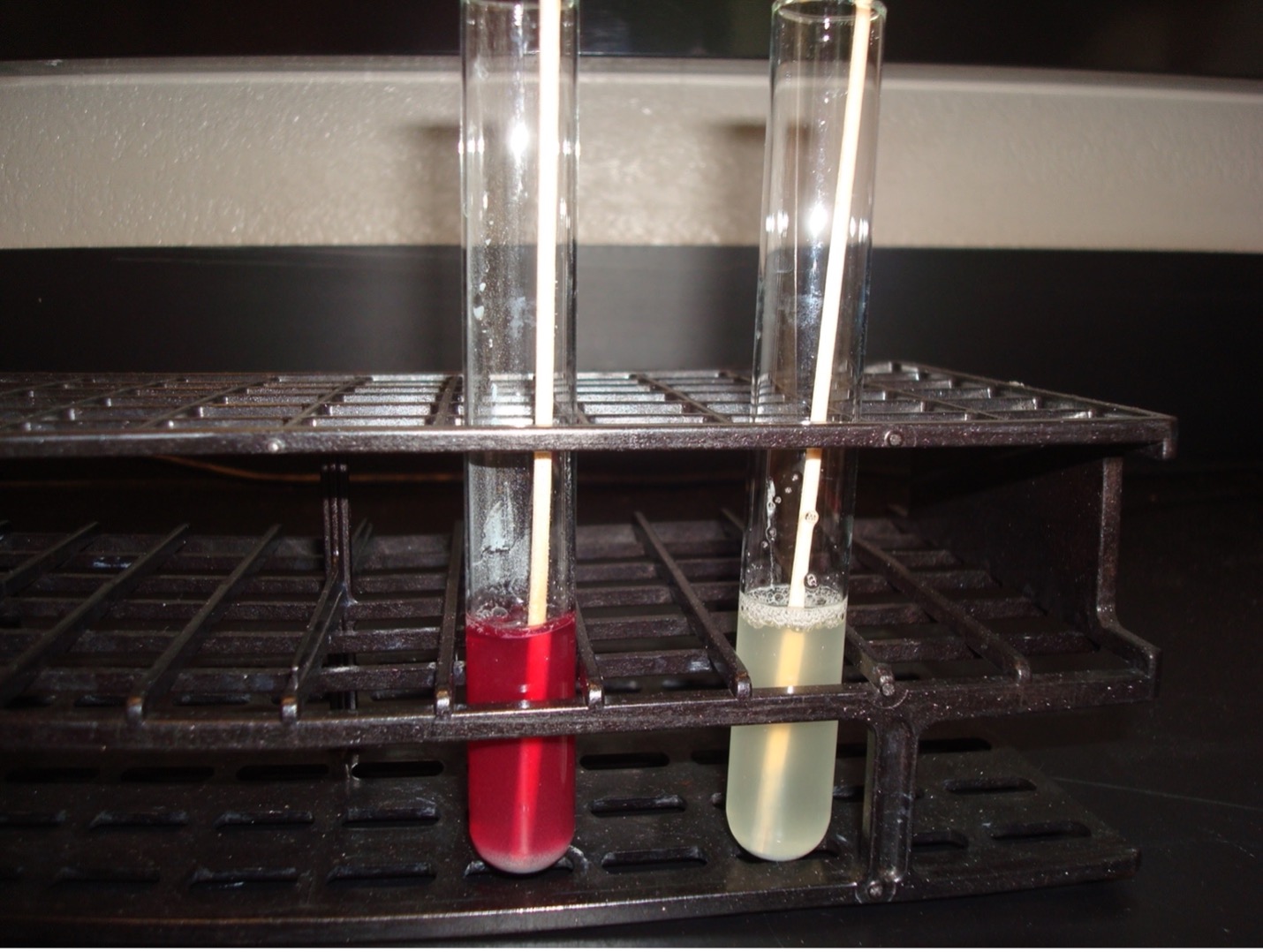
NITRATE REDUCTION TEST
Can the bacteria produce ATP via anaerobic energy production and use nitrate ad the final member of the electron transport chain? If it can use nitrate as the final member of the electron transport chain, does it reduce the nitrate to nitrite or nitrogen gas? Nitrate broth is a differential media used to determine if an organism can reduce nitrate. Some bacteria can reduce nitrate (NO3) to nitrite (NO2) by producing the enzyme nitrate reductase. Other bacteria can reduce nitrate to nitrogen gas by also producing the enzyme nitrite reductase which reduces nitrite to nitrogen gas. Other organisms do not have the ability to reduce nitrate at all.
Nitrate ——> Nitrite ——> Nitrogen Gas
AFTER INCUBATION
10 drops of 0.8% sulfanilic acid and then add 10 drops of 0.6% N, N-Dimethyl-alpha-naphthylamine are added to the incubated nitrate broth. A dark red color indicates the organism used the nitrate reductase enzyme to reduce nitrate to nitrite. This is a positive test result, and no further testing is required.
If no red color occurs a wooden applicator stick is dipped in zinc powder and dropped it into the nitrate broth. The zinc powder reduces nitrate to nitrite. A red color after the addition of zinc is a negative test result. Nitrate was not reduced by the organism, it was reduced by zinc. If a red color does not occur after the addition of zinc, there was no nitrate for zinc to reduce. This is considered a positive result since the organism reduced nitrate to nitrogen gas.
If no color, then add zinc
No color = Positive (Nitrate was reduced to Nitrogen gas)
Red color = Negative (Nitrate was not reduced by the bacteria)
identifying gram positive unknowns 1, 2, 3, and 4
Interpret the media and biochemical test results below for Gram Positive Unknowns 1, 2, 3, and 4 . Complete the data table and the identifications in the Identifying Gram Positive Unknowns Question Document.
UNKNOWN 1 MEDIA AND TEST RESULTS
UNKNOWN 2 MEDIA AND TEST RESULTS
UNKNOWN 3 MEDIA AND TEST RESULTS
UNKNOWN 4 MEDIA AND TEST RESULTS

DISCOVERIES IN MICROBIOLOGY
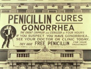 MARY HNATUSKO HUNT
In the 1940’s American bacteriologist Mary Hnatusko Hunt worked at a USDA research lab in Peoria Illinois. She visited the Illinois Fruit and Vegetable Company on Main Street in Peoria. She noticed the owner removing a moldy grapefruit from a display. She asked the owner if he could set aside moldy items for her to pick up on a regular basis, he agreed. At some point, Mary told him it was no longer necessary to save moldy fruit and vegetables for her. Several years later, Mary came to the store to inform the owner that a cantaloupe he provided was the source of penicillin. Mary helped isolate and test many strains of Penicillium.